Comparative Study
. 1997 Apr 29;94(9):4593-7.
doi: 10.1073/pnas.94.9.4593.

A novel chimeric Ig heavy chain from a teleost fish shares similarities to IgD

Affiliations
Comparative Study

A novel chimeric Ig heavy chain from a teleost fish shares similarities to IgD

M Wilson et al. Proc Natl Acad Sci U S A. .

Abstract

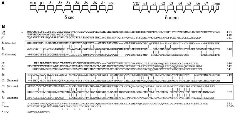
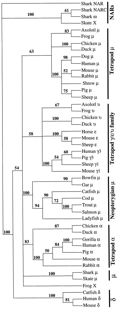
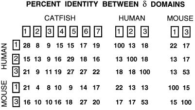
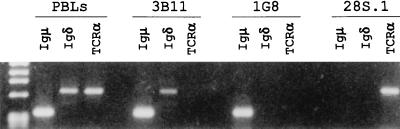
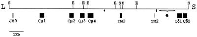
IgD is considered to be a recently evolved Ig, being previously found only in primates and rodents. Here we describe, from a teleost fish (the channel catfish, Ictalurus punctatus), a novel complex chimeric Ig heavy chain, homologous, in part, to the heavy chain (delta) of IgD. In addition to alternative secretory or membrane-associated C termini, this chimeric molecule contains a rearranged variable domain, the first constant domain of mu, and seven constant domains encoded by a delta gene homolog. Identification of the catfish gene as delta is based on the following properties: sequence relatedness to mammalian delta; a location within the IgH locus that is immediately downstream of the mu gene; separate terminal exons for the secretory and membrane forms; coexpression with the complete mu chain in some but not all B cells. These results (i) suggest that IgD is an ancient immunoglobulin that was present in vertebrates ancestral to both the mammals and the ray-finned fishes, and (ii) raise the possibility that this Ig isotype may have served an as yet unidentified important function early in the evolution of the immune system.


Figures

Figure 1
Figure 1
Structure and evolutionary relationships of the catfish IgD heavy chain. (A) Inferred exon structure encoding the secreted and membrane forms of the catfish δ chain. The exons are based on the deduced protein sequence of the Ig domain and on the identification of δ1 and δ2 exons in the germ line. (B) Complete amino acid sequence of the membrane form of catfish δ, inferred from the full-length cDNA M5 (accession no. U67437U67437). The sequence is shown by domain, and alignments with the homologous sequences of human and mouse δ C regions are boxed. The antigen receptor CART motif is marked with asterisks. The sequences of mouse δ are from ref. , and those of human δ are from ref. . The aligned regions (numbering of Kabat et al., ref. 28) are, for human δ1, 114–224; human δ2, 243–363; human δ3, 364–478; mouse δ1, 116–222; mouse δ3, 364–478. The sequence of the catfish δ secreted segment (δ sec) is taken from cDNA S1 (accession no. U67438U67438).
Figure 2
Figure 2
Phylogram of relationships between vertebrate Ig C regions. Sequences were aligned using clustal, with identity residue weight table, gap penalty 10, and gap-length penalty 10. The tree was generated from this alignment using paup (bootstrapping, with branch swapping/tree bisection-reconnection), and the values supporting each node are derived from 100 resamplings.
Figure 3
Figure 3
Comparison of the amino acid sequences of δ chains. Percent identity values from alignments of the individual domains (boxed) of catfish, mouse and human δ chains are shown.
Figure 4
Figure 4
Identification of catfish VH family usage in δ transcripts by RT-PCR. (a) RNA from freshly isolated PBLs was subjected to RT-PCR using forward primers specific for each of the seven described catfish VH families with a reverse primer at the 3′ end of the δ1 exon. The amplified products were electrophoresed on a 1% agarose gel and stained with ethidium bromide. The sizes of two HaeIII φX/174 DNA markers, 1078 and 872 (bp) are indicated by arrows. (b) Cμ1 sequences in the VH-δ1 PCR products were detected by blot hybridization analysis with a Cμ1 probe.
Figure 5
Figure 5
Expression of μ, δ, and TCRα mRNA in freshly-isolated PBLs, B cell lines 3B11 and 1G8, and a T cell line 28S.1. The RT-PCR products were electrophoresed on a 1% agarose gel. The markers (HaeIII φX/174 DNA) shown from top to bottom at left are: 1353, 1078, 872, 601, 281/271, and 234 bp. The VDJH rearrangements for both μ and δ have been sequenced for the B cell line 3B11 (accession nos. U67440U67440 and U67439U67439).
Figure 6
Figure 6
Physical map of the catfish IgH locus showing the μ gene and exons 1 and 2 of the δ gene. A partial restriction endonuclease map of recombinant phage 12C (35) is shown, with the exons encoding the first four catfish μ C region domains (Cμ1–4), the alternative membrane-anchoring C terminus of μ (TM1 and -2), and the first two δ exons (Cδ1 and -2) marked by boxes below the line. S and E are SalI and EcoRI sites, respectively. The L and S outside of the map indicate long and short phage arms of the EMBL 3 vector. The cleavage/polyadenylylation signal sequences are indicated by arrows, and the brackets mark the region of the enhancer, e (36). Accession number for the sequence of phage 12C is X79482X79482.

References

    1. DuPasquier L. In: Fundamental Immunology. 3rd Ed. Paul W E, editor. New York: Raven; 1993. pp. 199–223.
    1. Greenberg A S, Avila D, Hughes M, Hughes A, McKinney E C, Flajnik M F. Nature (London) 1995;374:168–173. - PubMed
    1. Greenberg A S, Hughes A L, Guo J, Avila D, McKinney E C, Flajnik M F. Eur J Immunol. 1996;26:1123–1129. - PubMed
    1. Bernstein R M, Schluter S F, Shen S, Marchalonis J J. Proc Natl Acad Sci USA. 1996;93:3289–3293. - PMC - PubMed
    1. Harding F A, Amemiya C T, Litman R T, Cohen N, Litman G W. Nucleic Acids Res. 1990;18:6369–6376. - PMC - PubMed

Publication types