Comparative Study
. 1997 Apr 29;94(9):4653-8.
doi: 10.1073/pnas.94.9.4653.

A new reporter cell line to monitor HIV infection and drug susceptibility in vitro

Affiliations
Comparative Study

A new reporter cell line to monitor HIV infection and drug susceptibility in vitro

A Gervaix et al. Proc Natl Acad Sci U S A. .

Abstract

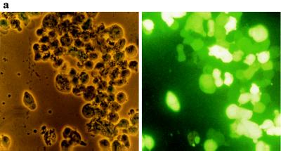
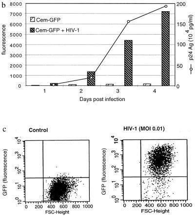
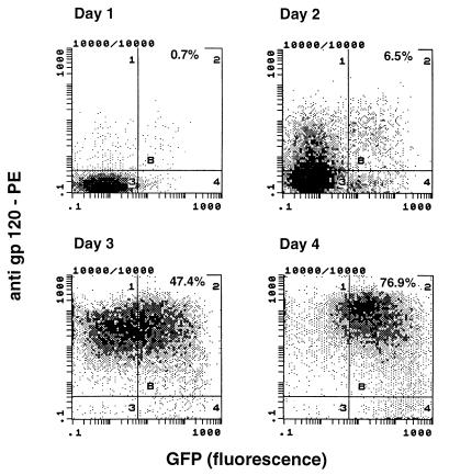
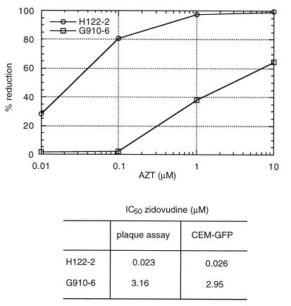
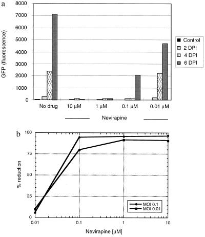
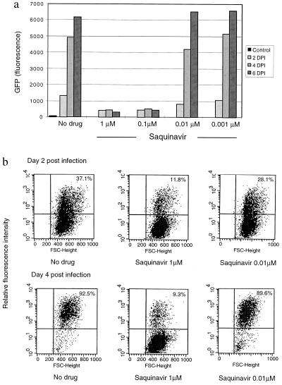
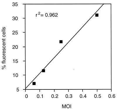
Determination of HIV infectivity in vitro and its inhibition by antiretroviral drugs by monitoring reduction of production of p24 antigen is expensive and time consuming. Such assays also do not allow accurate quantitation of the number of infected cells over time. To develop a simple, rapid, and direct method for monitoring HIV infection, we generated a stable T-cell line (CEM) containing a plasmid encoding the green fluorescent protein (humanized S65T GFP) driven by the HIV-1 long terminal repeat. Clones were selected that displayed low constitutive background fluorescence, but a high level of GFP expression upon infection with HIV. HIV-1 infection induced a 100- to 1,000-fold increase in relative fluorescence of cells over 2 to 4 days as monitored by fluorescence microscopy, cytofluorimetry, and flow cytometry. Addition of inhibitors of reverse transcriptase, protease, and other targets at different multiplicities of infection permitted the accurate determination of drug susceptibility. This technique also permitted quantitation of infectivity of viral preparations by assessment of number of cells infected in the first round of infection. In conclusion, the CEM-GFP reporter cell line provides a simple, rapid, and direct method for monitoring HIV infectivity titers and antiretroviral drug susceptibility of syncytium-inducing strains.


Figures

Figure 1
Figure 1
GFP expression in CEM-GFP cell line infected by HIV-1. (a) CEM-GFP cells 4 days after HIV-1 challenge at an input MOI of 0.01. CEM-GFP cells were visualized by conventional light-inverted microscopy (Left) and by fluorescence microscopy (Right). (b) Relative linear fluorescence quantification of 2 × 10⁵ CEM-GFP inoculated with HIV-1 at day 1, 2, 3, and 4 after infection (MOI 0.01). Cytofluorimetry was performed with an excitation wavelength of 485 nm and an emission wavelength of 508 nm, sensitivity 6. Parallel determination of the production of p24 antigen of infected cells by antigen capture ELISA test. (c) Analysis and enumeration of fluorescent CEM-GFP cells 4 days after infection with a MOI of 0.01 by flow cytometry.
Figure 1
Figure 1
GFP expression in CEM-GFP cell line infected by HIV-1. (a) CEM-GFP cells 4 days after HIV-1 challenge at an input MOI of 0.01. CEM-GFP cells were visualized by conventional light-inverted microscopy (Left) and by fluorescence microscopy (Right). (b) Relative linear fluorescence quantification of 2 × 10⁵ CEM-GFP inoculated with HIV-1 at day 1, 2, 3, and 4 after infection (MOI 0.01). Cytofluorimetry was performed with an excitation wavelength of 485 nm and an emission wavelength of 508 nm, sensitivity 6. Parallel determination of the production of p24 antigen of infected cells by antigen capture ELISA test. (c) Analysis and enumeration of fluorescent CEM-GFP cells 4 days after infection with a MOI of 0.01 by flow cytometry.
Figure 2
Figure 2
Comparative analysis of HIV-1 gp120 antigen and GFP expression in CEM-GFP cells infected with HIV-1 (MOI. 0.01). CEM-GFP inoculated with HIV-1 were incubated with mouse mAb against HIV-1 gp120 followed by a goat anti-mouse IgG antibody labeled with phycoerythrin at days 1 to 4 after infection. Cells were analyzed by flow cytometry gating for green (GFP) and red (anti-gp120).
Figure 3
Figure 3
Zidovudine susceptibility of the HIV-1 strains H112–2; zidovudine sensitive and its isogenic counterpart (G910–6) using cytofluorimetric measurements of infected cells obtained at days 4, 6 and 8 after infection (Upper). Cells were infected at a MOI of 0.1. The IC50 values as determined by plaque reduction in HeLa-CD4 cells and CEM-GFP assays are provided for comparison (Lower).
Figure 4
Figure 4
GFP expression of CEM-GFP challenged with HIV-1 at an input MOI of 0.1 in the presence of multiple concentrations (10 μM, 1 μM, 0.1 μM, and 0.01 μM) of the nonnucleoside reverse transcriptase inhibitor nevirapine. (a) Fluorescence measurement performed by cytofluorimetry (excitation wavelength 485 nm, emission wavelength 508 nm, sensitivity 6). DPI: days postinfection. (b) Determination of the IC50 of the nonnucleoside reverse transcriptase inhibitor nevirapine, using the results of a.
Figure 5
Figure 5
GFP expression of cells challenged with HIV in the presence of the protease inhibitor, saquinavir. (a) Fluorescence measurement by cytofluorimetry of CEM-GFP challenged with HIV-1 at an input MOI of 0.1 in the presence of multiple concentrations (1 μM, 0.1 μM, 0.01 μM, and 0.001 μM) of saquinavir. (b) HIV-challenged CEM-GFP cells incubated with 1 μM of saquinavir were also analyzed by flow cytometry. The percentage in the upper right quadrant indicates the proportion of cells infected in one round of infection.
Figure 6
Figure 6
Determination of viral infectivity titer. CEM-GFP were incubated at MOI of 0.06, 0.125, 0.25, and 0.5 with HIV-1LAI (titered on CEM cells by plaque assay) in the presence of 1 μM of saquinavir for 2 days. Cells were analyzed by flow cytometry.

References

    1. Richman D D, Johnson V A, Mayers D L, Shirasaka T, O’Brien M C, Mitsuya H. In: Current Protocols in Immunology. Ausubel F, Brent R, Kingston R E, Moore D D, Seidhan J G, Smith J A, Struhl K, editors. New York: Wiley; 1993. pp. 12.9.1–21.
    1. Hirsch M S, Kaplan J C. Sci Amer. 1987;256:76–85. - PubMed
    1. Schwartz O, Virelizier J L, Montagnier L, Hazan U. Gene. 1990;88:197–205. - PubMed
    1. Deng H, Liu R, Ellmeier W, Choe S, Unutmaz D, Burkhart M, Di Marzio P, Marmon S, Sutton R E, Mark Hill C, Davis C B, Peiper S C, Shall T J, Littman D R, Landau N R. Nature (London) 1996;381:661–666. - PubMed
    1. Merzouki A, Patel P, Cassol S, Ennaji M, Tailor P, Turcotte F R, O’Shaughnessy M, Arella M. Cell Mol Biol. 1995;41:445–452. - PubMed

Publication types

MeSH terms