. 1997 Apr 29;94(9):4659-63.
doi: 10.1073/pnas.94.9.4659.

A transgenic mouse model for measles virus infection of the brain

Affiliations

A transgenic mouse model for measles virus infection of the brain

G F Rall et al. Proc Natl Acad Sci U S A. .

Abstract

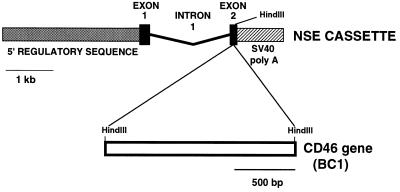
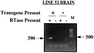
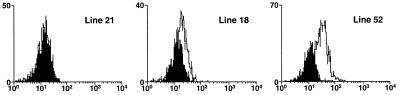
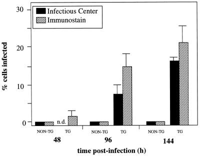
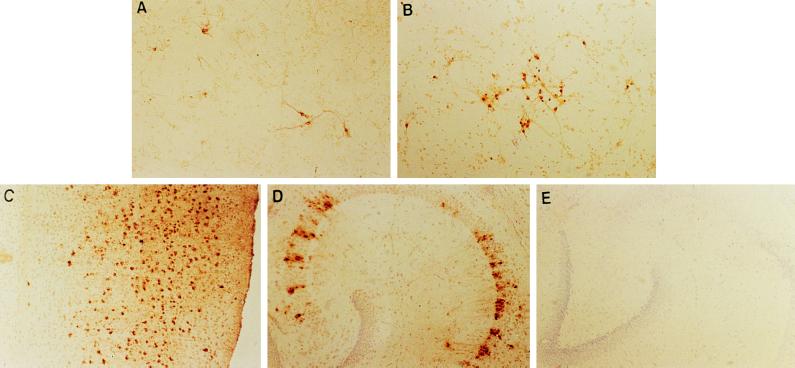
In addition to the rash, fever, and upper respiratory tract congestion that are the hallmarks of acute measles virus (MV) infection, invasion of the central nervous system (CNS) can occur, establishing a persistent infection primarily in neurons. The recent identification of the human membrane glycoprotein, CD46, as the MV receptor allowed for the establishment of transgenic mice in which the CD46 gene was transcriptionally regulated by a neuron-specific promoter. Expression of the measles receptor rendered primary CD46-positive neurons permissive to infection with MV-Edmonston. Notably, viral transmission within these cultures occurred in the absence of extracellular virus, presumably via neuronal processes. No infection was seen in nontransgenic mice inoculated intracerebrally with MV-Edmonston. In contrast, scattered neurons were infected following inoculation of transgenic adults, and an impressive widespread neuronal infection was established in transgenic neonates. The neonatal infection resulted in severe CNS disease by 3-4 weeks after infection. Illness was characterized initially by awkward gait and a lack of mobility, and in later stages seizures leading to death. These results show that expression of the MV receptor on specific murine cells (neurons) in vivo is absolutely essential to confer both susceptibility to infection and neurologic disease by this human virus. The disparity in clinical findings between neonatal and adult transgenic mice indicates that differences exist between the developing and mature CNS with respect to MV infection and pathogenesis.


Figures

Figure 1
Figure 1
Characterization of NSE–CD46 transgenic mice. The NSE–CD46 construct consists of 2.8 kb of 5′ NSE regulatory sequence, NSE exon 1 and intron 1, and 15 bp of NSE exon 2 adjacent to a unique HindIII site. Polyadenylylation signals are provided by the simian virus 40 sequence at the 3′ end of the construct, which also facilitates identification of transgenic mice by slot-blot analysis of genomic DNA.
Figure 2
Figure 2
Amplification of CD46 message in transgenic brains. Total RNA was isolated from four line 52 transgenic (+) and three nontransgenic (−) littermate brain homogenates, reverse transcribed and PCR amplified (5) using 20-mer oligonucleotides within the CD46 sequence. Samples were incubated with and without RT to verify amplification of a cDNA–RNA substrate. Representative results from one transgenic and one nontransgenic brain are shown.
Figure 3
Figure 3
Cell surface expression of CD46 on dissociated, transgenic neurons. Hippocampi from NSE–CD46 transgenic mouse lines 21, 18, and 52 were removed, dissociated, and incubated with the anti-CD46 monoclonal antibody E4.3 (19). The solid areas in each panel represent neurons from nontransgenic littermate controls stained using the identical procedure. Each panel depicts fluorescence intensity (x axis) vs. cell number (y axis). These results were subsequently confirmed with two additional hippocampal preparations.
Figure 4
Figure 4
Detection of MV in primary neuron cultures. Primary neurons cultured from transgenic (TG) (line 52) and nontransgenic (NON-TG) embryonic hippocampi were infected with MV–Edmonston at a multiplicity of infection = 3, 48 hr after culturing. At various times after infection, neurons were harvested for infectious center analysis or immunostained with SSPE serum. Supernatants were also collected for plaque assay. For the immunostaining, 10 fields per time point were counted, and standard deviations are shown.
Figure 5
Figure 5
Spread of MV in primary neurons and in vivo. (A) CD46+ primary neurons, infected for 48 hr with MV–Edmonston. (B) CD46+ primary neurons 96 hr after infection. Coverslips were fixed with 1:1 acetone:methanol and immunostained with a human SSPE serum as described. (CE) NSE–CD46 neonatal (<24 postnatal) transgenic and nontransgenic mice were inoculated intracranially with 105 pfu of MV–Edmonston and brain sections were immunostained for MV as described. Immunohistochemical staining for MV antigens in the cortical region (C) and in the hippocampus (D) of a representative, NSE–CD46 transgenic mouse infected with MV 10 days previously (×100). (E) Hippocampus of a nontransgenic mouse, inoculated 10 days previously (×100).

References

    1. Norrby E, Oxman M. In: Virology. Fields B N, Knipe D M, editors. New York: Raven; 1990. pp. 1013–1045.
    1. Griffin D E, Bellini W J. In: Virology. Fields B N, Knipe D M, Howley P M, editors. Philadelphia: Lippincott–Raven; 1996. pp. 1267–1312.
    1. ter Meulen V, Stephenson J R, Kreth H W. Compr Virol. 1983;8:105–159.
    1. Katayama Y, Hotta H, Nishimura A, Tatsuno Y, Homma M. J Gen Virol. 1995;76:3201–3204. - PubMed
    1. Liebert U G, Finke D. Curr Top Microbiol Immunol. 1995;191:149–166. - PubMed

Publication types

MeSH terms