. 1997 Apr 29;94(9):4675-80.
doi: 10.1073/pnas.94.9.4675.

A proline-rich motif in p53 is required for transactivation-independent growth arrest as induced by Gas1

Affiliations

A proline-rich motif in p53 is required for transactivation-independent growth arrest as induced by Gas1

E M Ruaro et al. Proc Natl Acad Sci U S A. .

Abstract

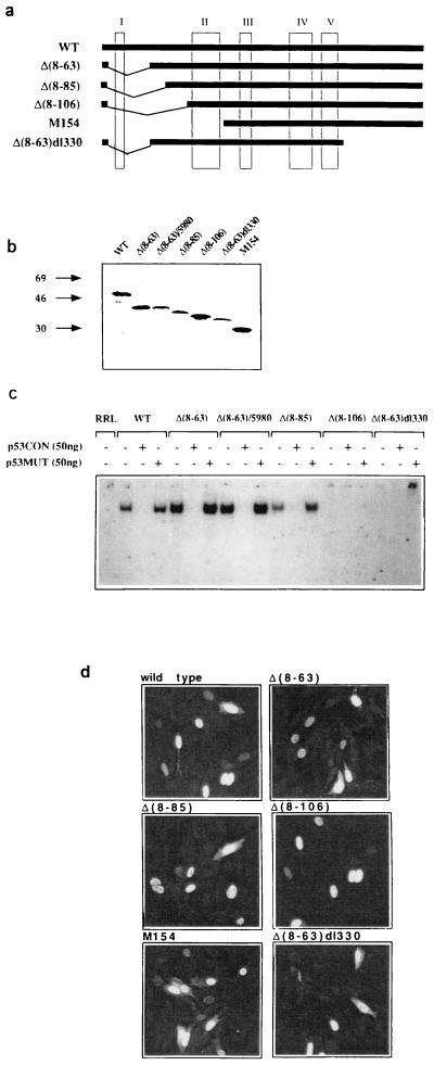
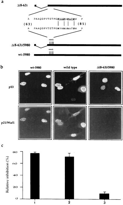
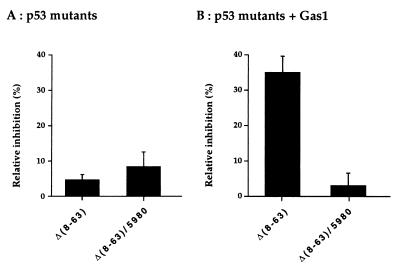
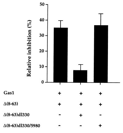
The involvement of p53 in regulating diverse cellular processes dictates that it must respond to multiple signaling mechanisms, thus coordinating the response to various "stress conditions." Genotoxic stress has served as a paradigm to dissect the transactivation-dependent branch of the pathway by which p53 can induce growth arrest. Alternate mechanisms have been invoked to explain transactivation-independent effects of p53, especially in the context of apoptosis. We have identified a p53-dependent pathway initiated by the gas1 product, a plasma membrane protein highly expressed during G0, which activates a transactivation-independent p53 growth arrest function. Through a detailed deletional analysis and site-specific mutagenesis of p53 we show that the Gas1-dependent signal transduction relies on a proline-rich region (amino acids 63-85) of murine p53. In vivo competition experiments using combinations of such mutants implicate this functional domain of p53 as a docking site in the transmission of antiproliferative signals.


Figures

Figure 1
Figure 1
Expression, sequence specific DNA binding activity, and nuclear localization of murine p53 deletion mutants. (a) Schematic representation of the p53 mutants used. Numbering of murine p53 amino acids is done according to ref. . (b) In vitro translated murine p53 mutants were analyzed by SDS/PAGE to confirm the expected apparent molecular weights. The structure of deletion Δ(8–63)/5980 is described in Fig. 3. (c) In vitro translated proteins were tested for sequence specific DNA binding by electrophoretic mobility shift assay with the p53CON oligonucleotide as a probe. Specificity of the binding was confirmed by competition with excess cold p53CON or p53MUT oligonucleotides. (d) 100 ng/μl of each pGDSV7 expression plasmid (31) containing the different p53 deletion mutants, was microinjected into the nuclei of BALB/c (10)1 cells (36). p53 protein expression was analyzed 12 hr later by indirect immunofluorescence using PAb240. Images were obtained with a LSM 450 confocal microscope (Zeiss).
Figure 2
Figure 2
Growth arrest analysis of the p53 deletion mutants in BALB/c (10)1 fibroblasts. (A) Relative inhibition of BrdUrd incorporation by expression of the various p53 mutants. (B) Relative inhibition of BrdUrd incorporation after coexpression of the various p53 mutants in combination with Gas1. pGDVS7 expression vector (100 ng/μl of each) was microinjected into the nuclei of growing BALB/c (10)1 fibroblasts. When Gas1 was expressed together with the p53 deletion mutants, 50 ng/μl of each plasmid was used. Eighteen hours after injection 50 μM BrdUrd was added to the culture medium to monitor S phase. After 6 hr cells were fixed and processed for immunofluorescence as described in Materials and Methods. The percentage of relative inhibition of DNA synthesis in the injected cells was calculated by the following formula: % relative inhibition = [% BrdUrd-positive cells (uninjected) − % of BrdUrd-positive cells (p53/Gas1-positive)/% of BrdUrd-positive cells (uninjected)]. The mean of three independent experiments with at least 300 overexpressing cells scored is shown.
Figure 3
Figure 3
Structure, p21/Waf1 induction and growth arrest function analysis of proline-rich region mutants. (a) Amino acid sequence of the proline-rich region in mouse p53. The PXXP motifs are underlined; the substituted amino acids in the mutated version are indicated in bold letters. Clone p53wt-5980 codes for a murine p53 bearing four point mutations that substitute Pro-76, -79, and -81 with alanines, and Pro-84 with serine. (b) Proline substitution in the p53 sequence does not interfere with its ability to transactivate endogenous p21/Waf1 as assayed by the induction of the corresponding protein product. Expression plasmids were microinjected in BALB/c (10)1 cells. Six hours later cells were fixed and analyzed for expression of the p53 protein and of endogenous p21/Waf1 by immunofluorescence. p21/Waf1 was detected by anti-peptide antibody as described (29). Wt p53 and Δ(8–63)/5980 were used as positive and negative controls, respectively. (c) Analysis of the growth arrest function of p53 wt-5980 (1), wt p53 (2), and Δ(8–3)/5980 (3). BrdUrd incorporation was monitored as described in legend to Fig. 2.
Figure 4
Figure 4
Mutation of Pro-76, -79, -81, and -84 within deletion mutant p53 Δ(8–63) affects the ability to reconstitute the Gas1-dependent growth arrest function. (A) Relative inhibition of BrdUrd incorporation by expression of Δ(8–63) or Δ(8–63)/5980 p53 mutants. (B) Relative inhibition of BrdUrd incorporation by co-expression of the same p53 mutants in combination with Gas1. Microinjection experiments were performed as described in legend to Fig. 2. The mean of three independent experiments with at least 300 overexpressing cells scored are shown.
Figure 5
Figure 5
Model for Gas1 signaling via the proline-rich domain of p53. The proline-rich domain of p53 (dots indicate the four relevant prolines) may signal for growth suppression via contacting the SH3 domain of a cellular protein (X). Such interaction should be modulated by signals departing from the plasma membrane where Gas1 is localized. A p53 mutant lacking the tetramerization domain (competitor) could interfere with growth suppression by sequestering the cognate SH3-containing cellular protein.
Figure 6
Figure 6
Triple co-expression of Gas1 plus Δ(8–63) and either Δ(8–63)dl330 or Δ(8–63)dl330/5980 deletion mutants in BALB/c (10)1 cells. Δ(8–63)dl330 mutant, that retains the proline-rich domain but lacks the transactivation and oligomerization domains, behaves as an efficient competitor for the Gas1/Δ(8–63) inhibitory effect. Competition is not observed with the corresponding proline-substituted mutant Δ(8–63)dl330/5980. Microinjection experiments were performed as described in legend to Fig. 2. pGDSV7-gas1 (50 ng/μl) plus 30 ng/μl of pGDSV7-Δ(8–63), and 30 ng/μl of either pGDSV7-Δ(8–63)dl330 or pGDSV7-Δ(8–63)dl330/5980 were microinjected into BALB/c (10)1 cells. Experiments were scored either for Gas1 or p53 expression relative to BrdUrd incorporation giving similar results. Mean ± SD.

References

    1. Levine A J. Annu Rev Biochem. 1993;63:623–651. - PubMed
    1. Bates S, Vousden K H. Curr Opin Gen Dev. 1996;6:12–19. - PubMed
    1. Gottlieb T M, Oren M. Biochem Biophys Acta Rev Cancer. 1996;1287:77–102. - PubMed
    1. Ko L J, Prives C. Genes Dev. 1996;9:1054–1072. - PubMed
    1. Graeber T G, Peterson J F, Tsai M, Monica K, Fornace A J, Jr, Giaccia A J. Mol Cell Biol. 1994;14:6264–6277. - PMC - PubMed

Publication types