Comparative Study
. 1997 Apr 29;94(9):4727-32.
doi: 10.1073/pnas.94.9.4727.

Localization of guanylate cyclase-activating protein 2 in mammalian retinas

Affiliations
Comparative Study

Localization of guanylate cyclase-activating protein 2 in mammalian retinas

A Otto-Bruc et al. Proc Natl Acad Sci U S A. .

Abstract

Guanylate cyclase-activating proteins (GCAP1 and GCAP2) are thought to mediate the intracellular stimulation of guanylate cyclase (GC) by Ca2+, a key event in recovery of the dark state of rod photoreceptors after exposure to light. GCAP1 has been localized to rod and cone outer segments, the sites of phototransduction, and to photoreceptor synaptic terminals and some cone somata. We used in situ hybridization and immunocytochemistry to localize GCAP2 in human, monkey, and bovine retinas. In human and monkey retinas, the most intense immunolabeling with anti-GCAP2 antibodies was in the cone inner segments, somata, and synaptic terminals and, to a lesser degree, in rod inner segments and inner retinal neurons. In bovine retina, the most intense immunolabeling was in the rod inner segments, with weaker labeling of cone myoids, somata, and synapses. By using a GCAP2-specific antibody in enzymatic assays, we confirmed that GCAP1 but not GCAP2 is the major component that stimulates GC in bovine rod outer segment homogenates. These results suggest that although GCAP1 is involved in the Ca2+-sensitive regulation of GC in rod and cone outer segments, GCAP2 may have non-phototransduction functions in photoreceptors and inner retinal neurons.


Figures

Figure 1
Figure 1
In situ hybridization of GCAP2 transcripts in the human retina using antisense (A) and sense (B) probes. The following layers are indicated: OS, photoreceptor outer segments; IS, photoreceptor inner segments; ONL, outer nuclear layer; OPL, outer plexiform layer; INL, inner nuclear layer; IPL, inner plexiform layer; GCL, ganglion cell layer; and NFL, nerve-fiber layer. The strongest labeling with the antisense probe is in the cone inner segments (arrows), with weaker labeling of the rod inner segments (arrowheads). (Bar = 50 μm.)
Figure 2
Figure 2
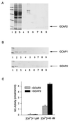
Immunoaffinity chromatography of retinal extracts. The GCAP1 and GCAP2 were extracted from 50 retinas as described by Gorczyca et al. (17). The extract was loaded onto an immunoaffinity column containing affinity-purified anti-GCAP2 polyclonal antibodies (UW50) coupled to CNBr-activated Sepharose. The fractions that passed through the column during loading were collected, and the column was washed. Bound proteins were eluted with 0.1 M glycine (pH 2.5). Eluted fractions were immediately neutralized with 1 M Tris⋅HCl (pH 8.4). (A) SDS/PAGE analysis. The indicated fractions (in 15 μl) were analyzed on 12.3% polyacrylamide gels and stained with Coomassie brilliant blue R-250. Lanes: 1, standard proteins of known molecular masses (from the top in kDa: 92, 67, 43, 30, 21, 14; from Pharmacia); 2, extracts before chromatography; 3, fractions that passed through the column; 4, fractions that passed through the column in the BTP buffer; 5, fractions that passed through the column in 4 M urea; 6–9, fractions eluted with glycine (pH 2.5). The arrow indicates the position of GCAP2. (B) Reactivity of retinal extracts with anti-GCAP1 and anti-GCAP2 antibodies. One-microliter aliquots from fractions as described in A were probed with anti-GCAP1 monoclonal antibody (G-2) or with anti-GCAP2 polyclonal antibodies (UW31). The arrows indicate the positions of GCAP1 and GCAP2, respectively. Note that G-2 antibody does not react with GCAP2 on immunoblots, although at the high concentrations of G-2 coupled to the CNBr-activated Sepharose, GCAP2 was retained on the column [discussed by Gorczyca et al. (17)]. (C) GC-stimulating activity of GCAP2. Ten-microliter aliquots of a fraction with the highest concentration of GCAP2 was tested to determine its effect on GC activity with washed ROS membranes in the presence of either 1 μM Ca2+ or 45 nM Ca2+.
Figure 3
Figure 3
Immunofluorescence localization of GCAP2 in monkey retina. (A) GCAP2 immunolabeling with UW50 is strongest in the cone inner segments (arrows), somata (arrowheads), and synaptic terminals. Cone nuclei are negative images. Weak immunolabeling is present in the rod and cone outer segments (denoted by asterisk), rod inner segments, and neurons in the inner retina. (B) Although individual cones are rarely visible in their entirety within a single image plane, their cellular processes can be visualized with a projection of a z series. Arrows denote cone synaptic terminals. (C) Addition of purified GCAP1 (25 μg/ml) to anti-GCAP2 polyclonal antibodies produces minimal decrease in GCAP2 immunoreactivity, verifying that this antibody does not cross-react with GCAP1. (D) Addition of GCAP2–7 (25 μg/ml) to anti-GCAP2 polyclonal antibodies abolishes GCAP2 immunoreactivity. Sections incubated in preimmune serum (E) or buffer without anti-GCAP2 (F) show no immunolabeling of cones or other retinal cell types. The choroid (at the top) is weakly reactive with the secondary antibody. (Bar = 20 μm.)
Figure 4
Figure 4
Immunofluorescence localization of GCAP2 and cone opsins in monkey retina. (A–C) Localization of GCAP2 and red/green cone opsin. (A) The cones are immunolabeled with anti-GCAP2 (UW50), with the strongest labeling in the inner segments, somata, and synaptic terminals. Some inner retinal neurons are weakly labeled. (B) Anti-red/green cone opsin labels the majority of cone outer segments. (C) Double labeling with anti-red/green cone opsin (green) and anti-GCAP2 (red) demonstrates that the red/green cones are immunopositive for GCAP2. (D–F) Localization of GCAP2 and blue cone opsin. (D) The cone photoreceptors are immunolabeled with anti-GCAP2. (E) Anti-blue cone opsin labels a single cone outer segment. (F) Double labeling with anti-blue cone opsin (green) and anti-GCAP2 (red) demonstrates that the blue cone is immunopositive for GCAP2. (Bar = 20 μm.)
Figure 5
Figure 5
Immunofluorescence localization of GCAP2 in bovine retina. (A) Both rod and cone photoreceptors are labeled. In rods, labeling is strongest in the inner segments (asterisk). The cone inner segments are substantially wider than rod inner segments and the cell bodies of cones are restricted to the outermost tier of the outer nuclear layer. Labeling is present in the myoid region of a cone inner segment (arrowhead), cone somata (arrows) and a cone axon (a). Labeled photoreceptor synapses are at the bottom. (Bar = 15 μm.) (B) Addition of GCAP2–7 (25 μg/ml) to anti-GCAP2 polyclonal antibodies abolishes GCAP2 immunoreactivity.
Figure 6
Figure 6
Inhibition of GC activity in ROS. (A) SDS/PAGE and immunoblots of GCAP1 and GCAP2 isolated from a retinal extract. UW14 was specific for GCAP1, and UW31 was specific for GCAP2. S represents molecular markers at 20 kDa. Note that GCAP2 appears in two molecular forms of the unknown origin and that UW31 does not recognize GCAP1. (B) The inhibition of GC activity in ROS homogenates. The GC assay was performed as described by Gorczyca et al. (4). Dotted line was determined experimentally from the inhibition of GCAP2-dependent stimulation of GC in reconstituted systems composed of purified GCAP2 (2 μg) and washed ROS membranes. Solid line was determined experimentally from the inhibition of GCAP1-dependent stimulation of GC in reconstituted systems composed of purified GCAP1 (6 μg) and washed ROS membranes.The amount of GCAP1 or GCAP2 (studied in the range of GC stimulation between 5 and 80 pmol/min) did not affect the slope (GC activity versus antibody concentration) of these inhibitions.

Similar articles

Cited by

References

    1. Koutalos Y, Yau K-W. Trends Neurosci. 1996;19:73–81. - PubMed
    1. Polans A, Baehr W, Palczewski K. Trends Neurosci. 1996;19:547–554. - PubMed
    1. Koch K-W, Stryer L. Nature. 1988;334:64–66. - PubMed
    1. Gorczyca W A, Gray-Keller M P, Detwiler P B, Palczewski K. Proc Natl Acad Sci USA. 1994;91:4014–4018. - PMC - PubMed
    1. Dizhoor A M, Lowe D G, Olshevskaya E V, Laura R P, Hurley J B. Neuron. 1994;12:1345–1352. - PubMed

Publication types

MeSH terms

Substances

LinkOut - more resources