. 1997 Apr 29;94(9):4739-44.
doi: 10.1073/pnas.94.9.4739.

The regulator of early gliogenesis glial cells missing is a transcription factor with a novel type of DNA-binding domain

Affiliations

The regulator of early gliogenesis glial cells missing is a transcription factor with a novel type of DNA-binding domain

J Schreiber et al. Proc Natl Acad Sci U S A. .

Abstract

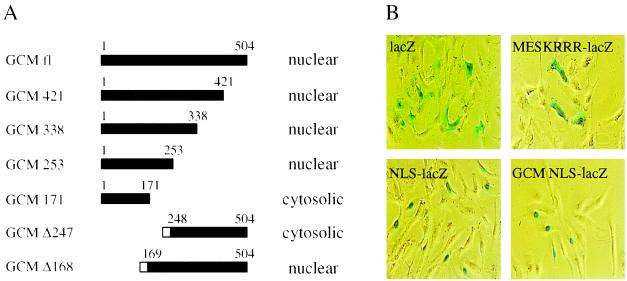
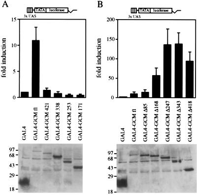
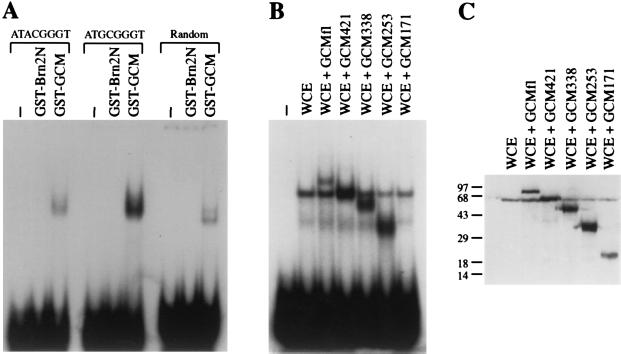
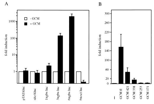
Absence or presence of glial cells missing (GCM) in cells of the developing nervous system of Drosophila decides over their future fate as neurons or glia with only those cells turning into glia that express GCM. To understand how GCM exerts its function we performed a detailed structure-function analysis. Using fusions between the DNA binding domain of the yeast GAL4 protein and GCM, we detected a transactivation function within the C-terminal part of GCM. In addition to this transactivation domain we mapped a sequence-specific DNA-binding domain within the N-terminal part of the GCM protein in close proximity to a bipartite nuclear localization signal. Binding site selection assays determined the motif 5'-AT(G/A)CGGGT-3' as the preferred binding site for GCM. Both the lack of homology to known proteins and the novel DNA binding specificity indicate that GCM contained a new type of DNA-binding domain. In transiently transfected cells, GCM also activated transcription from promoters consisting of the newly identified GCM-binding site and a TATA box. Thus, GCM is a novel type of transcription factor involved in early gliogenesis.


Figures

Figure 1
Figure 1
Nuclear localization of GCM. (A) Cellular distribution of full-length GCM and various mutants as detected in immunofluorescence studies on transiently transfected CV1 cells. Mutants GCM 421, GCM 338, GCM 253, and GCM 171 as well as full-length GCM were detected using rabbit anti-GCM antiserum, whereas the tagged mutants GCM Δ247 and GCM Δ168 were visualized using a mAb directed against the T7-tag (Novagen). Amino acids present within each mutant are indicated by the filled bar; the open box represents the T7-tag. (B) Localization of β-gal by histochemical X-gal staining in CV1 cells transiently transfected with pCMVlacZ (lacZ), pCMVlacZ/Tst-1 NLS (NLS-lacZ), pCMV/MESKRRRlacZ (MESKRRR-lacZ), or pCMVlacZ/GCM NLS (GCM NLS-lacZ).
Figure 2
Figure 2
Transactivation potential of GCM. A GAL4-responsive luciferase reporter (3xUAS TATA luciferase) was transfected into U138 glioblastoma cells together with expression plasmids for the GAL4 DNA-binding domain (GAL4) or for fusions of the GAL4 DNA-binding domain with full-length GCM (GAL4–GCM fl) or mutant versions thereof. (A) Fusions containing C-terminal deletion mutants of GCM: GAL4–GCM 421, GAL4–GCM 338, GAL4–GCM 253, GAL4–GCM 171. (B) Fusions containing N-terminal deletion mutants of GCM: GAL4–GCM Δ85, GAL4–GCM Δ168, GAL4–GCM Δ247, GAL4–GCM Δ343, GAL4–GCM Δ418. (Upper) Luciferase activities in extracts from transfected cells were determined in four independent experiments, each performed in duplicates. Data are presented for each GAL4–GCM fusion as fold induction above the level of luciferase activity obtained in transfections with an expression plasmid for the GAL4 DNA-binding domain, which was given an arbitrary value of 1. (Lower) Expression of GAL4 fusion proteins in transfected cells was confirmed by Western blot analyses of whole cell extracts. Numbers on left indicate size of molecular weight markers in kDa.
Figure 3
Figure 3
DNA-binding of GCM. (A) Purified GST–GCM and GST–Brn2N protein were analyzed in electrophoretic mobility shift assays for their ability to bind to radiolabeled oligonucleotides containing either the sequence 5′-ATGCGGGT-3′ or 5′-ATACGGGT-3′, or to a random oligonucleotide which had undergone three of six selection cycles for an optimal GCM-binding site. (B) The radiolabeled oligonucleotide containing the 5′-ATGCGGGT-3′ motif was used to detect complex formation with whole cell extracts from COS cells that were either mock-transfected (WCE), transfected with full length GCM (WCE + GCM fl) or transfected with various GCM mutants (WCE + GCM 421, WCE + GCM 338, WCE + GCM 253, WCE + GCM 171). (C) Western blot analyses of cell extracts used in B. Numbers on left indicate size of molecular weight markers in kDa.
Figure 4
Figure 4
In vivo function of GCM. (A) Various luciferase reporter plasmids were transfected into U138 glioblastoma cells in the absence (open bars) or presence (filled bars) of cotransfected full-length GCM. pTATAluc contained only a minimal promoter, whereas all other reporter plasmids carried additional binding sites for transcription factors. siteAluc and 6xoct luc contained one or six binding sites for POU-domain proteins; gbs luc constructs had either one, three, or six copies of a GCM-binding site inserted in front of the minimal promoter. (B) The 3xgbs luc reporter was transfected together with empty expression plasmid (−) or various GCM expression plasmids (GCM fl, GCM 421, GCM 338, GCM 253, and GCM 171) into U138 cells. Luciferase activities in extracts from transfected cells were determined in three independent experiments, each performed in duplicate. Data are presented as fold inductions that were calculated for each reporter plasmid by comparing luciferase activities to values from cells that were transfected with reporter plasmid and empty CMV expression plasmid. Note the logarithmic scale of the y axis in A.

References

    1. Anderson D J. Neuron. 1989;3:1–12. - PubMed
    1. Doe C Q, Technau G M. Trends Neurosci. 1993;16:510–514. - PubMed
    1. Jan Y N, Jan L Y. Curr Opin Neurobiol. 1994;4:8–13. - PubMed
    1. Hosoya T, Takizawa K, Nitta K, Hotta Y. Cell. 1995;82:1025–1036. - PubMed
    1. Jones B W, Fetter R D, Tear G, Goodman C S. Cell. 1995;82:1013–1023. - PubMed

Publication types

MeSH terms

LinkOut - more resources