

Cell death prevention, mitogen-activated protein kinase stimulation, and increased sulfatide concentrations in Schwann cells and oligodendrocytes by prosaposin and prosaptides
- PMID: 9114068
- PMCID: PMC20801
- DOI: 10.1073/pnas.94.9.4778
Cell death prevention, mitogen-activated protein kinase stimulation, and increased sulfatide concentrations in Schwann cells and oligodendrocytes by prosaposin and prosaptides
Abstract
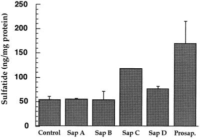
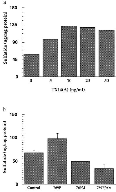
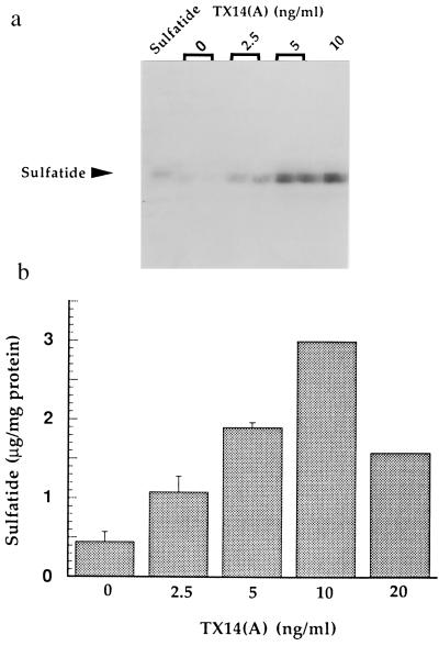
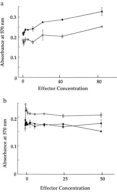
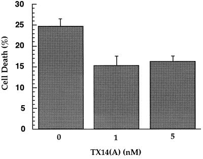
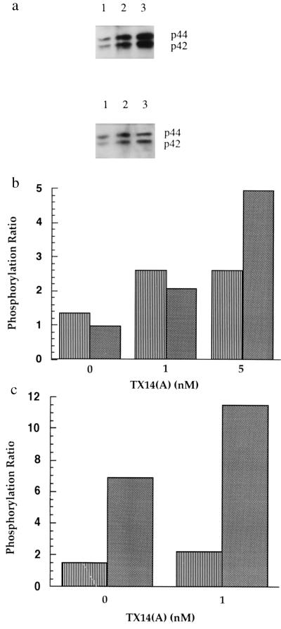
Prosaposin, the precursor of saposins A, B, C, and D, was recently identified as a neurotrophic factor. Herein prosaposin was found to increase sulfatide concentrations in primary and transformed Schwann cells (iSC) and oligodendrocytes (differentiated CG4 cells). Of the four mature saposins, only saposin C was found to increase sulfatide concentrations in these cell types. A similar result was obtained by using peptides (prosaptides) encompassing the neurotrophic sequence located in the saposin C domain. Dose-response curves demonstrated maximal enhancement by saposin C and prosaptides at low nanomolar concentrations (5-10 nM). The increase in sulfatide concentration by a 14-mer prosaptide, TX14(A), in CG4 oligodendrocytes was about 3-fold greater than in primary Schwann cells. A mutant prosaptide with a single amino acid replacement of Asn --> Asp was inactive. Prosaptides did not induce cell proliferation of primary Schwann cells, iSC cells, or CG4 oligodendrocytes but nanomolar concentrations of prosaptides prevented cell death of iSC cells and CG4 oligodendrocytes. Immunoblot analysis demonstrated that phosphorylation of both mitogen-activated protein kinase p-42 and p-44 isoforms were enhanced 3- to 5-fold after 5 min of treatment with prosaptides at concentrations of 1-5 nM. These findings suggest that prosaposin and prosaptides bind to a receptor that initiates signal transduction to promote myelin lipid synthesis and prolong cell survival in both Schwann cells and oligodendrocytes. Prosaposin may function as a myelinotrophic factor in vivo during development and repair of myelinated nerves explaining the deficiency of myelin observed in prosaposin-deficient mice and humans.
Figures
References
-
- O’Brien J S, Kishimoto Y. FASEB J. 1991;5:301–308. - PubMed
-
- O’Brien J S, Carson G S, Seo H-C, Hiraiwa M, Weiler S, Tomich J M, Barranger J A, Kahn M, Azuma N, Kishimoto Y. FASEB J. 1995;9:681–685. - PubMed
-
- Campana W M, Hiraiwa M, Addison K C, O’Brien J S. Biochem Biophys Res Commun. 1996;229:706–712. - PubMed
-
- Sano A, Matsuda S, Wen T-C, Kotani Y, Kondoh K, Ueno S, Kakimoto Y, Yoshimura H, Sakanaka M. Biochem Biophys Res Commum. 1994;204:994–1000. - PubMed
Publication types
MeSH terms
Substances
Grants and funding
LinkOut - more resources
Full Text Sources
Other Literature Sources
Miscellaneous
