. 1997 Apr 29;94(9):4782-7.
doi: 10.1073/pnas.94.9.4782.

Insulin secretion is regulated by the glucose-dependent production of islet beta cell macrophage migration inhibitory factor

Affiliations

Insulin secretion is regulated by the glucose-dependent production of islet beta cell macrophage migration inhibitory factor

G Waeber et al. Proc Natl Acad Sci U S A. .

Abstract

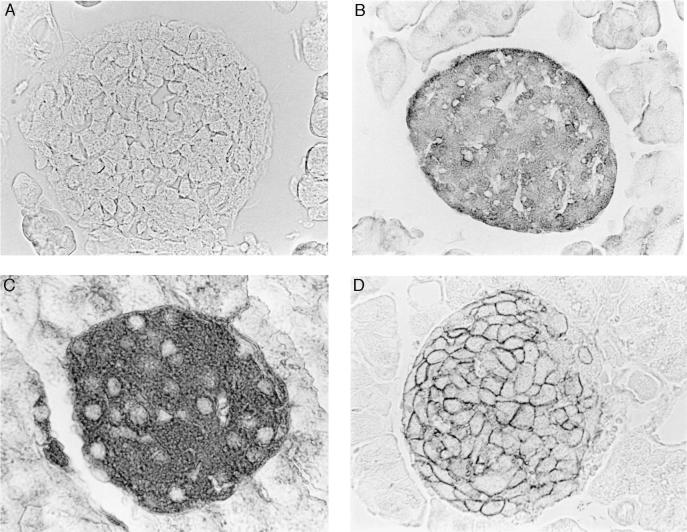
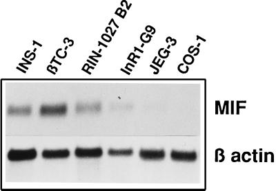
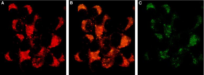
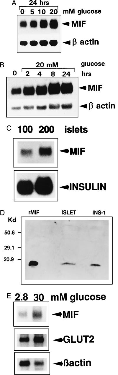
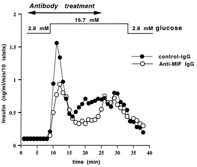
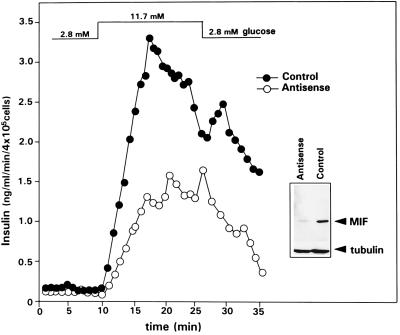
Macrophage migration inhibitory factor (MIF), originally identified as a cytokine secreted by T lymphocytes, was found recently to be both a pituitary hormone and a mediator released by immune cells in response to glucocorticoid stimulation. We report here that the insulin-secreting beta cell of the islets of Langerhans expresses MIF and that its production is regulated by glucose in a time- and concentration-dependent manner. MIF and insulin colocalize by immunocytochemistry within the secretory granules of the pancreatic islet beta cells, and once released, MIF appears to regulate insulin release in an autocrine fashion. In perifusion studies performed with isolated rat islets, immunoneutralization of MIF reduced the first and second phase of the glucose-induced insulin secretion response by 39% and 31%, respectively. Conversely, exogenously added recombinant MIF was found to potentiate insulin release. Constitutive expression of MIF antisense RNA in the insulin-secreting INS-1 cell line inhibited MIF protein synthesis and decreased significantly glucose-induced insulin release. MIF is therefore a glucose-dependent, islet cell product that regulates insulin secretion in a positive manner and may play an important role in carbohydrate metabolism.


Figures

Figure 1
Figure 1
Immunohistochemical localization of MIF in the islets of Langerhans of the pancreas. MIF is detected within the cytoplasm of islet cells (B) that also are positive for insulin (C) and GLUT2 (D). The GLUT2-positive staining is localized predominantly to cell membranes (10, 13). No MIF is detected when cells are stained with preimmune serum (negative control) (A).
Figure 2
Figure 2
MIF is highly expressed in pancreatic β cell lines. Northern blotting analyses of MIF mRNA (0.6 kb) and β-actin mRNA (1.8 kb) expression in two insulin-producing cell lines (INS-1 and βTC3), an insulin and somatostatin cell line (RIN1027-B2), a glucagon-producing cell line (InR1-G9), and nonpancreatic cell lines (JEG-3 and COS-1). Total RNA (10 μg) was prepared and analyzed as described in Methods. The MIF transcript is detected in high abundance in the insulin-producing cell lines.
Figure 3
Figure 3
MIF is colocalized in the insulin-containing granule of β cells. INS-1 cells were stained with anti-insulin (A), anti-insulin and anti-MIF (B), or anti-MIF IgG (C) and revealed by immunofluorescence (Texas Red for the anti-insulin antibodies and fluorescein isothiocyanate for the anti-MIF). Insulin granules are red, MIF staining is green, and the orange-yellow staining corresponds to granules containing MIF and insulin. Fluorescence images were obtained with confocal laser scan microscope. (×60.)
Figure 4
Figure 4
Expression and glucose-dependent regulation of MIF mRNA in pancreatic β cell lines and in isolated rat pancreatic islets. (A) Northern blot analysis of MIF mRNA and β-actin mRNA expression by INS-1 cells incubated with 0, 5, 10, and 20 mM glucose in RPMI 1640 medium for 24 hr. (B) Time course of MIF mRNA expression by INS-1 cells incubated with 20 mM glucose. Total RNA was extracted at intervals, and MIF mRNA was analyzed by Northern blotting as described in the Methods. (C) Islets of Langerhans were isolated from Sprague–Dawley rats, total RNA was extracted from 100 and 200 islets and analyzed by Northern blotting using MIF and rat proinsulin II cDNA probes. (D) Western blot analysis of the MIF content of INS-1 cells or isolated islets of Langerhans. Protein extracts (40 μg) obtained from INS-1 cells or islets of Langerhans were size-fractionated on an SDS/18% polyacrylamide gel, transferred to nitrocellulose, and blotted with an anti-MIF antiserum. rMIF (50 ng) served as a positive control and size marker. (E) Northern blot analysis of MIF, GLUT-2, and β-actin mRNA expression by 400 pancreatic islets incubated for 12 hr in RPMI 1640 medium containing 2.8 or 30 mM glucose. Normalized to β-actin, MIF and GLUT2 mRNA increased 4-fold when the glucose concentration was raised from 2.8 to 30 mM.
Figure 5
Figure 5
Immunoneutralization of islet cell MIF inhibits insulin secretion. Anti-MIF antibodies decrease the first and second phase of glucose-induced insulin secretion by isolated pancreatic islets. Ten purified rat islets were incubated in a perifusion chamber and the glucose concentration of the perifused buffer was increased experimentally from 2.8 mM (−40 to +7 min) to 16.7 mM (+8 to +33 min) and then returned to 2.8 mM (+34 min) as shown. The results are expressed as the mean ± SEM of three separate perifusion experiments. Control, nonimmune IgG or anti-MIF IgG (each at 50 μg/ml) was added to the chamber from +2 to +24 min. The quantity of insulin released during the first phase (+8 to +15 min) and the second phase (+16 to +25 min) of insulin secretion showed a decrease after immunoneutralization of MIF from 1.17 ± 0.19 ng/ml to 0.71 ± 0.06 ng/ml and 0.74 ± 0.02 ng/ml to 0.51 ± 0.01 ng/ml, respectively (P < 0.01 and P < 0.001 for the first and second phases of insulin secretion, respectively; Student’s t test).
Figure 6
Figure 6
MIF antisense RNA inhibits MIF protein expression and glucose-induced insulin release by INS-1 cells. Antisense MIF or parental (control) expression vector were stably integrated into INS-1 cells. Western blotting of the MIF content of the cell lysates (20 μg total protein) obtained from an antisense transfected clone versus a control clone showed a significant decrease in the MIF protein content in cells containing the antisense construct (Inset). Control or antisense INS-1 cells then were incubated in a perifusion chamber, and the glucose concentration of the perifused buffer (supplemented with 1 mM 3-isobutyl-1-methyl-xanthine and 1 μM forskolin) increased from 2.8 mM (−40 to +8 min) to 11.7 mM (+9 to +26 min) and then returned to 2.8 mM (+27 min).

References

    1. Bloom B R, Bennet B. Science. 1966;153:80–82. - PubMed
    1. David J. Proc Natl Acad Sci USA. 1966;56:72–77. - PMC - PubMed
    1. Bernhagen J, Calandra T, Mitchell R A, Martin S B, Tracey K J, Voelter W, Manogue K R, Cerami A, Bucala R. Nature (London) 1993;365:756–759. - PubMed
    1. Calandra T, Bernhagen J, Mitchell R A, Bucala R. J Exp Med. 1994;179:1895–1902. - PMC - PubMed
    1. Nishino T, Bernhagen J, Shiki H, Calandra T, Dohi K, Bucala R. Mol Med. 1995;1:781–788. - PMC - PubMed

Publication types