Comparative Study
. 1997 Apr 29;94(9):4794-9.
doi: 10.1073/pnas.94.9.4794.

A membrane-anchored E-type endo-1,4-beta-glucanase is localized on Golgi and plasma membranes of higher plants

Affiliations
Comparative Study

A membrane-anchored E-type endo-1,4-beta-glucanase is localized on Golgi and plasma membranes of higher plants

D A Brummell et al. Proc Natl Acad Sci U S A. .

Abstract

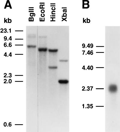
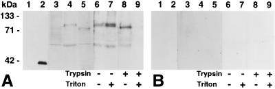
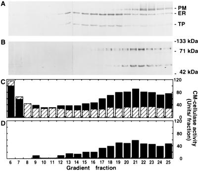
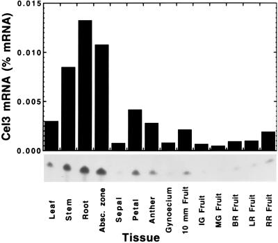
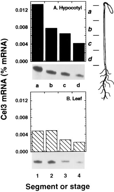
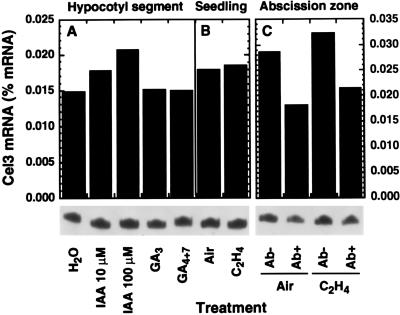
Endo-1,4-beta-D-glucanases (EGases, EC 3.2.1.4) are enzymes produced in bacteria, fungi, and plants that hydrolyze polysaccharides possessing a 1,4-beta-D-glucan backbone. All previously identified plant EGases are E-type endoglucanases that possess signal sequences for endoplasmic reticulum entry and are secreted to the cell wall. Here we report the characterization of a novel E-type plant EGase (tomato Cel3) with a hydrophobic transmembrane domain and structure typical of type II integral membrane proteins. The predicted protein is composed of 617 amino acids and possesses seven potential sites for N-glycosylation. Cel3 mRNA accumulates in young vegetative tissues with highest abundance during periods of rapid cell expansion, but is not hormonally regulated. Antibodies raised to a recombinant Cel3 protein specifically recognized three proteins, with apparent molecular masses of 93, 88, and 53 kDa, in tomato root microsomal membranes separated by sucrose density centrifugation. The 53-kDa protein comigrated in the gradient with plasma membrane markers, the 88-kDa protein with Golgi membrane markers, and the 93-kDa protein with markers for both Golgi and plasma membranes. EGase enzyme activity was also found in regions of the density gradient corresponding to both Golgi and plasma membranes, suggesting that Cel3 EGase resides in both membrane systems, the sites of cell wall polymer biosynthesis. The in vivo function of Cel3 is not known, but the only other known membrane-anchored EGase is present in Agrobacterium tumefaciens where it is required for cellulose biosynthesis.


Figures

Figure 1
Figure 1
Sequence and phylogenetic analysis. (A) Deduced amino acid sequence of tomato Cel3 aligned with tomato Cel1 and Cel2 mature proteins. Amino acid identity is indicated by asterisks and conserved changes by dots. Potential N-glycosylation sequences in Cel3 are overlined. (B) Schematic representation of tomato Cel3 protein structure. Cel3 consists of a charged N terminus (shaded box), a transmembrane domain (solid box), and the catalytic core (hatched box). (C) Hydropathic profile of Cel3 deduced protein. (D) Phylogenetic comparison of plant EGase deduced proteins after removal of signal sequences. Sequences and GenBank accession numbers: avocado, Cel1 (M17634M17634); bean, BAC1 (M57400M57400); elder, JET1 (X74290X74290); pea, EGL1 (L41046L41046); peach, Cel1 (X96853X96853); pepper, Cel1 (X87323X87323), Cel2 (X97190X97190), and Cel3 (X97189X97189); poplar, Cel1 (D32166D32166); tomato, Cel1 (U13054U13054), Cel2 (U13055U13055), and Cel4 (also called TPP18; U20590U20590).
Figure 2
Figure 2
DNA and RNA gel blot analysis. (A) DNA gel blot of 10-μg aliquots of genomic DNA digested with the indicated restriction enzyme. The gel blot was hybridized with a 32P-labeled 1117-nucleotide Cel3 cDNA fragment in 50% formamide, 0.9 M NaCl (6× SSPE) at 36°C, with final wash in 0.075 M NaCl (0.5× SSC) at 53°C (27°C below Tm). (B) RNA gel blot of 3 μg of stem poly(A)+ RNA hybridized with an antisense Cel3 riboprobe in 50% formamide, 0.75 M NaCl (5× SSPE) at 59°C, with final wash in 0.015 M NaCl (0.1× SSC) at 49°C (23°C below Tm).
Figure 3
Figure 3
Western blots of uninduced and induced E. coli proteins (100 ng per lane) and tomato proteins (50 μg per lane) probed with Cel3 antibody (1:7,500) affinity-purified against recombinant Cel3 protein blotted to transfer membrane (A) or against transfer membrane alone blocked with BSA (B). Lanes: 1, uninduced E. coli; 2, E. coli induced with isopropyl β-d-thiogalactoside; 3, tomato soluble proteins; 4, tomato microsomal membranes; 5, tomato cell wall proteins; 6–9, microsomal membranes treated with no trypsin, no Triton (lane 6), no trypsin, 0.1% Triton (lane 7); 500 units of trypsin, no Triton (lane 8); 500 units of trypsin, 0.1% Triton (lane 9).
Figure 4
Figure 4
Distribution of membrane marker enzymes in a sucrose density gradient used to separate tomato root microsomal membranes. (A) Total protein determined using Bradford assay and sucrose concentration measured by refractometry. (B) NADH:cytochrome c reductase (ER) and vanadate-sensitive ATPase (plasma membrane). (C) Bafilomycin-sensitive ATPase (tonoplast) and latent UDPase (Golgi). Marker enzyme and protein assays performed as described (13).
Figure 5
Figure 5
Western blots of tomato root microsomal membrane density gradient fractions probed with antibodies to plasma membrane ATPase (100 kDa), ER-localized BiP (70 kDa), and 58-kDa subunit of tonoplast ATPase (58 kDa) (A); or affinity-purified Cel3 antibody (B). (C) CM-cellulase activity determined using viscometry in the absence (striped bars) or presence (solid bars) of 0.1% Triton X-100. (D) Triton-activated CM-cellulase activity calculated from C.
Figure 6
Figure 6
Cel3 mRNA abundance in various tomato vegetative, flower, and fruit tissues. Cel3 mRNA abundance was determined against a standard curve using ribonuclease protection assays.
Figure 7
Figure 7
Cel3 mRNA abundance in young expanding tissues. (A) Zones of etiolated hypocotyl, as indicated in the Inset. (B) Expansion of young green leaves. Stages: 1, 1.0 × 0.3 cm; 2, 2.5 × 1.0 cm; 3, 3.5 × 1.5 cm; 4, 5.5 × 3.0 cm.
Figure 8
Figure 8
Effect of hormone treatments on Cel3 mRNA abundance. (A) Etiolated hypocotyl segments incubated for 24 h in solutions of IAA, 10 μM GA3, or 10 μM GA4+7. (B) Young etiolated seedlings exposed to air or 10 μl/l ethylene for 24 h. (C) Abscission zones of flower explants treated with air or 10 μl/l ethylene, then nonabscised (Ab−) and abscised (Ab+) zones were collected separately.

Similar articles

Cited by

References

    1. Beguin P. Annu Rev Microbiol. 1990;44:219–248. - PubMed
    1. Brummell D A, Lashbrook C C, Bennett A B. Am Chem Soc Symp Ser. 1994;566:100–129.
    1. Koehler D E, Leonard R T, Vanderwoude W J, Linkins A E, Lewis L N. Plant Physiol. 1976;58:324–330. - PMC - PubMed
    1. del Campillo E, Durbin M, Lewis L N. Plant Physiol. 1988;88:904–909. - PMC - PubMed
    1. Lewis L N, Koehler D E. Planta. 1979;146:1–5. - PubMed

Publication types

MeSH terms

Associated data

LinkOut - more resources