. 1997 Mar 3;185(5):805-16.
doi: 10.1084/jem.185.5.805.

C-C chemokines released by lipopolysaccharide (LPS)-stimulated human macrophages suppress HIV-1 infection in both macrophages and T cells

Affiliations

C-C chemokines released by lipopolysaccharide (LPS)-stimulated human macrophages suppress HIV-1 infection in both macrophages and T cells

A Verani et al. J Exp Med. .

Abstract

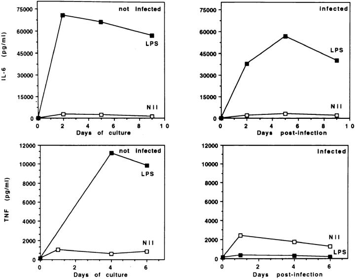
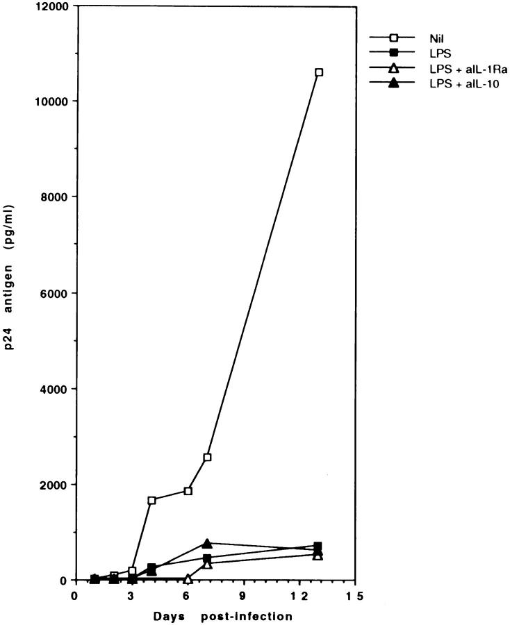
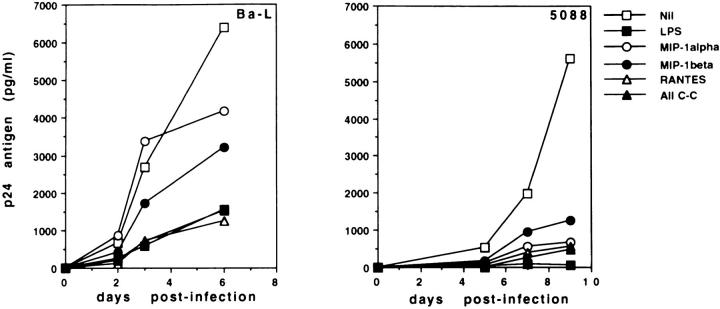
Human immunodeficiency virus-1 (HIV-1) expression in monocyte-derived macrophages (MDM) infected in vitro is known to be inhibited by lipopolysaccharide (LPS). However, the mechanisms are incompletely understood. We show here that HIV-1 suppression is mediated by soluble factors released by MDM stimulated with physiologically significant concentrations of LPS. LPS-conditioned supernatants from MDM inhibited HIV-1 replication in both MDM and T cells. Depletion of C-C chemokines (RANTES, MIP-1 alpha, and MIP-1 beta) neutralized the ability of LPS-conditioned supernatants to inhibit HIV-1 replication in MDM. A combination of recombinant C-C chemokines blocked HIV-1 infection as effectively as LPS. Here, we report an inhibitory effect of C-C chemokines on HIV replication in primary macrophages. Our results raise the possibility that monocytes may play a dual role in HIV infection: while representing a reservoir for the virus, they may contribute to the containment of the infection by releasing factors that suppress HIV replication not only in monocytes but also in T lymphocytes.


Figures

Figure 6
Figure 6
MDM express CC–CKR-5 mRNA. Total RNA was extracted from untreated MDM. RNA samples were treated with DNase I to remove traces of contaminating DNA and reverse transcribed using random hexameric primers. The cDNA products were mixed to scalar amounts of a synthetic competitor DNA fragment containing primer recognition sites for both β-actin and CC–CKR-5 amplification, and amplified with the respective primer pairs. (A) Schematic representation of the competitor DNA fragment used for the quantification of CC–CKR-5 and β-actin cDNA. The fragment contains a core sequence derived from the human β-actin cDNA, carrying a 20-bp insertion in the middle (closed box). Amplification with the β-actin-specific primer set BA1–BA4 detects a 226-bp product on human cDNA, and a 246-bp product from the competitor DNA. To this core sequence, the primer recognition sites for human CC–CKR-5 amplification were added at the two ends (indicated by gray boxes) by reamplification with composite primers corresponding to the CKR-9+BA1 sequence at one end and CKR-10+BA4 at the other end. Amplification with CKR-9 and CKR-10 generates a 288-bp fragment from the competitor template and a 368-bp fragment from the CC–CKR-5 cDNA. (B) Competitive PCR for the quantification of CC– CKR-5 and β-actin mRNAs. cDNA samples from untreated MDM were mixed with tenfold dilution of the competitor DNA fragment as indicated, and amplified with primer sets CKR-9/CKR-10 and BA1/BA4 for CC–CKR-5 and β-actin mRNA quantification. Amplification products were resolved by polyacrylamide gel electrophoresis, stained with ethidium bromide, and quantified by densitometric scanning. According to the principles of competitive PCR, quantification of the target molecules in the samples was obtained by estimation of the ratio between the amplification products, as reported at the bottom of each gel. Furthermore, since the same competitor DNA fragment acts as a competitor for quantification of both CC–CKR-5 and β-actin, standardization for mRNA input is obtained by estimating the ratio between the two measurements, as indicated at the bottom of the figure. M, molecular weight markers.
Figure 1
Figure 1
LPS suppresses HIV-1 replication in MDM cultures infected in vitro. MDM from healthy donors were infected with HIV-1Ba-L (A), the primary NSI isolate HIV-15088 (B), or HIV-1IIIB (D), all at 500 pg/ml, in the presence or absence of LPS (1 μg/ml). MDM were washed 1 d later and further cultured, adding LPS every 3 d. Culture supernatants were harvested daily, and tested for p24 Ag secretion by ELISA. The data are representative of 10 (A), 3 (B), and 2 (D) separate experiments. In (C) MDM were infected with HIV-1Ba-L or HIV-15088 in the presence of decreasing concentrations of LPS. p24 Ag secretion was assessed 5 d after infection.
Figure 2
Figure 2
LPS-induced inhibition of HIV-1 expression in MDM cultures is dependent on the time of addition of LPS. MDM were infected with HIV-1Ba-L (500 pg/ml), and stimulated with LPS (1 μg/ml) at different times from the initiation of the culture. Culture supernatants were harvested daily, and tested for p24 Ag secretion by ELISA. The data represent the mean of two separate experiments.
Figure 3
Figure 3
LPS and HIV-1 synergize in upregulating CD14 expression in MDM. MDM were infected with HIV-1Ba-L (500 pg/ml) in the presence or absence of LPS (1 μg/ml). After 2 d of culture, CD14 expression was assessed by direct immunofluorescence, using PE-conjugated mAb P9 and an unrelated isotype control. The data are representative of three separate experiments.
Figure 4
Figure 4
Effects of LPS stimulation and/or HIV-1 infection on IL-6 and TNF-α secretion by MDM. Uninfected or HIV-1Ba-Linfected MDM were cultured in the presence or absence of LPS (1 μg/ml). LPS was added to the cultures every 3 d. IL-6 and TNF-α concentrations in the supernatants were measured by ELISA. The data are representative of four separate experiments.
Figure 5
Figure 5
Effects of neutralizing antibodies against HIV-1-inhibitory cytokines. MDM were infected with HIV-1Ba-L and stimulated with LPS (1 μg/ml), in presence or absence of neutralizing anti-IL-1Ra or anti-IL-10 antibodies (10 μg/ml). Culture supernatants were harvested daily, and tested for p24 Ag secretion by ELISA. The data represent the mean of two separate experiments. Control antibodies had no effect on p24 Ag secretion.
Figure 7
Figure 7
Recombinant C–C chemokines inhibit HIV-1 replication in human MDM. MDM from healthy donors were infected in vitro with HIV-1Ba-L (left) or with the NSI primary viral isolate HIV-15088 (right), in the presence or absence of LPS (1 μg/ml) and recombinant chemokines. Chemokines were added to HIV-1Ba-L-infected cultures at a concentration of 250 ng/ml when used individually, and 50 ng/ml each when used in combination. For HIV-15088infected cultures, chemokines were used at 10 ng/ml, individually and in combination. Supernatants from infected cultures were harvested at different timepoints, and assayed by ELISA for p24 Ag secretion.

References

    1. Stoler MH, Eskin TA, Benn S, Angerer RC, Angerer LM. Human T-cell lymphotrophic virus type III infection of the central nervous system. Preliminary in situ analysis. JAMA. 1986;256:2360–2364. - PubMed
    1. Koenig S, Gendelman HE, Orenstein JM, Canto MCD, Pezeshkpour GM, Yungbluth M, Janotta F, Aksamit A, Martin MA, Fauci AS. Detection of AIDS virus in macrophages in brain tissue from AIDS patients with encephalopathy. Science (Wash DC) 1986;233:1089–1093. - PubMed
    1. Gendelman HE, Orenstein JM, Martin MA, Ferrua C, Mitra R, Phipps T, Wahl LA, Lane HC, Fauci AS, Burke DS, et al. Efficient isolation and propagation of human immunodeficiency virus on recombinant colony-stimulating factor 1–treated monocytes. J Exp Med. 1988;167:1428–1441. - PMC - PubMed
    1. Weiss SH, Goedert JJ, Gartner S, Popovic M, Waters D, Markham P, Di Marzo F, Veronese, Gail MH, Barkley WE, Gibbons J, et al. Risk of human immunodeficiency virus infection among laboratory workers. Science (Wash DC) 1988;239:68–71. - PubMed
    1. Ho DD, Rota TR, Hirsh MS. Infection of monocytes/macrophages by human T lymphotropic virus type III. J Clin Invest. 1986;77:1712–1715. - PMC - PubMed

Publication types

MeSH terms