Comparative Study
. 1997 Mar 3;185(5):817-24.
doi: 10.1084/jem.185.5.817.

Regulation of the interleukin (IL)-12R beta 2 subunit expression in developing T helper 1 (Th1) and Th2 cells

Affiliations
Comparative Study

Regulation of the interleukin (IL)-12R beta 2 subunit expression in developing T helper 1 (Th1) and Th2 cells

S J Szabo et al. J Exp Med. .

Abstract

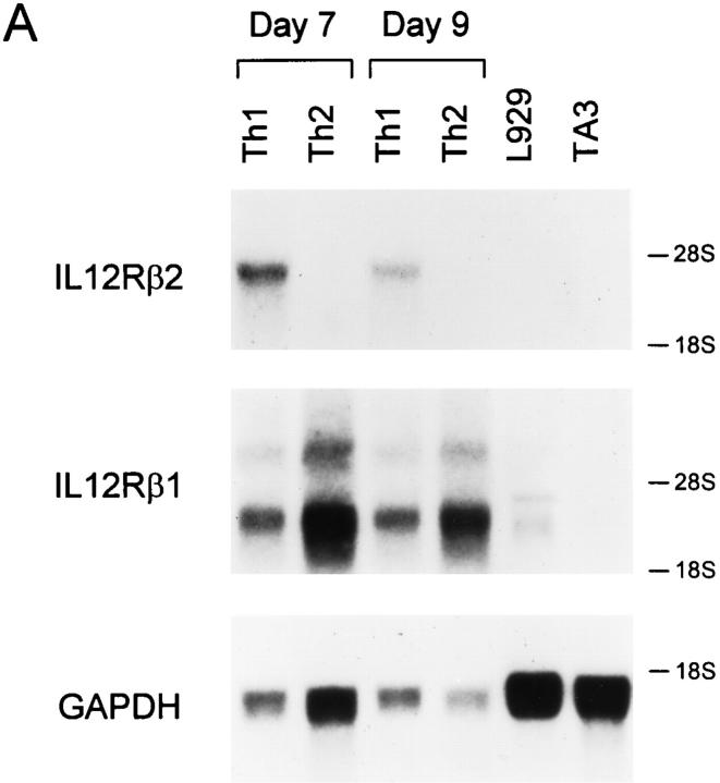
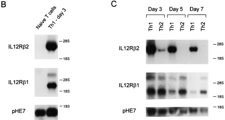
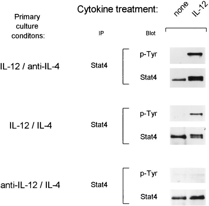
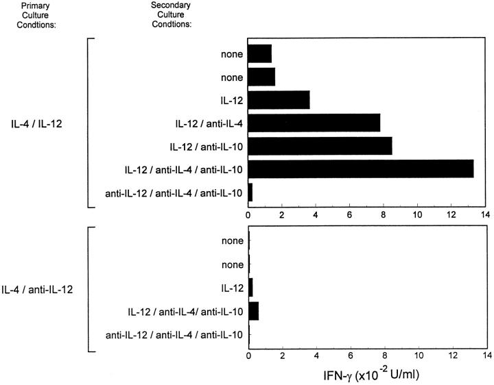
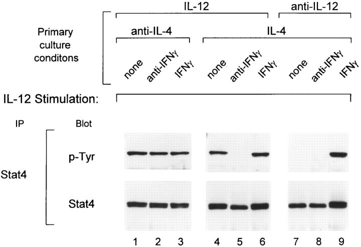
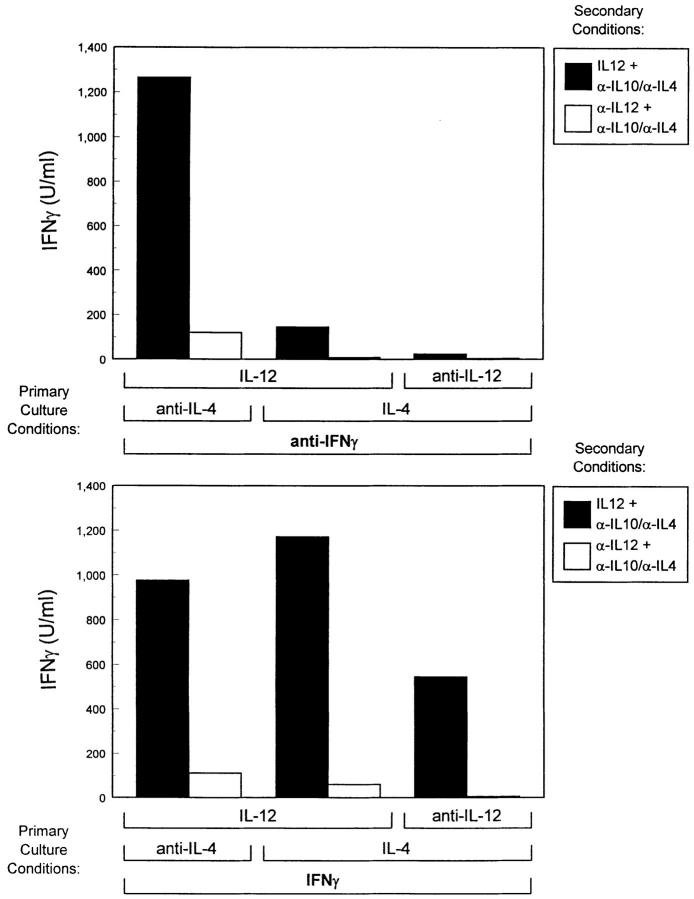
The developmental commitment to a T helper 1 (Th1)- or Th2-type response can significantly influence host immunity to pathogens. Extinction of the IL-12 signaling pathway during early Th2 development provides a mechanism that allows stable phenotype commitment. In this report we demonstrate that extinction of IL-12 signaling in early Th2 cells results from a selective loss of IL-12 receptor (IL-12R) beta 2 subunit expression. To determine the basis for this selective loss, we examined IL-12R beta 2 subunit expression during Th cell development in response to T cell treatment with different cytokines. IL-12R beta 2 is not expressed by naive resting CD4+ T cells, but is induced upon antigen activation through the T cell receptor. Importantly, IL-4 and IFN-gamma were found to significantly modify IL-12 receptor beta 2 expression after T cell activation. IL-4 inhibited IL-12R beta 2 expression leading to the loss of IL-12 signaling, providing an important point of regulation to promote commitment to the Th2 pathway. IFN-gamma treatment of early developing Th2 cells maintained IL-12R beta 2 expression and restored the ability of these cells to functionally respond to IL-12, but did not directly inhibit IL-4 or induce IFN-gamma production. Thus, IFN-gamma may prevent early Th cells from premature commitment to the Th2 pathway. Controlling the expression of the IL-12R beta 2 subunit could be an important therapeutic target for the redirection of ongoing Th cell responses.


Figures

Figure 1
Figure 1
IL-12R β2 subunit mRNA is detected in Th1 but not Th2 cells. (A) Naive CD4+ T cells were purified by FACS® from unimmunized DO11.10 TCR-transgenic mice as described in Materials and Methods (5), activated with OVA peptide and APCs under either Th1- or Th2-inducing conditions and allowed to develop for 7 days. On day 7, the Th1 and Th2 cells were washed, restimulated and allowed to proliferate for 7 or 9 d when cells were harvested and total cellular RNA was isolated. As tissue controls, total cellular RNA was isolated from the B cell hybridoma TA3 and the fibroblast cell line L929. Northern blot analysis was performed using as probes the full-length murine IL-12R β2 subunit cDNA (top), the full-length murine IL-12R β1 subunit cDNA (middle), and the GAPDH cDNA (bottom). (B and C). Total cellular RNA was isolated from naive T cells after purification by FACS®. Naive T cells isolated by cell sorting were activated to induce Th1 or Th2 development, and harvested on days 3, 5, and 7 after primary antigen activation. Total cellular RNA was examined by Northern analysis as described above for IL-12R β2 subunit cDNA (top), the IL-12R β1 subunit cDNA (middle), or pHE7 cDNA (bottom).
Figure 1
Figure 1
IL-12R β2 subunit mRNA is detected in Th1 but not Th2 cells. (A) Naive CD4+ T cells were purified by FACS® from unimmunized DO11.10 TCR-transgenic mice as described in Materials and Methods (5), activated with OVA peptide and APCs under either Th1- or Th2-inducing conditions and allowed to develop for 7 days. On day 7, the Th1 and Th2 cells were washed, restimulated and allowed to proliferate for 7 or 9 d when cells were harvested and total cellular RNA was isolated. As tissue controls, total cellular RNA was isolated from the B cell hybridoma TA3 and the fibroblast cell line L929. Northern blot analysis was performed using as probes the full-length murine IL-12R β2 subunit cDNA (top), the full-length murine IL-12R β1 subunit cDNA (middle), and the GAPDH cDNA (bottom). (B and C). Total cellular RNA was isolated from naive T cells after purification by FACS®. Naive T cells isolated by cell sorting were activated to induce Th1 or Th2 development, and harvested on days 3, 5, and 7 after primary antigen activation. Total cellular RNA was examined by Northern analysis as described above for IL-12R β2 subunit cDNA (top), the IL-12R β1 subunit cDNA (middle), or pHE7 cDNA (bottom).
Figure 2
Figure 2
Activation in the presence of IL-4 and IL-12 results in the maintenance of the IL-12 signaling pathway. FACS®-sorted naive CD4+ DO11.10 T cells were cultured with OVA peptide and irradiated BALB/c splenocytes in the presence of 10 U/ml IL-12 and 10 μg/ml anti–IL-4 to promote Th1 differentiation (upper), 10 U/ml IL-12 and 200 U/ml IL-4 (middle), or 200 U/ml IL-4 and 3 μg/ml anti–IL-12 to promote Th2 differentiation (bottom). After 72 h the cells were expanded 10-fold in fresh medium containing 40 U/ml IL-2. On day 5 after primary antigen activation whole cell lysates from developing T cells (1.5–2.0 × 10⁷) were prepared after incubation for 25 min with medium alone or with 10 U/ml IL-12. Lysates were immunoprecipitated with Stat4 antiserum, separated by SDS-PAGE (7% gel), transferred to nitrocellulose, and probed with the anti-phosphotyrosine reagent RC20 as described (30). After exposure, blots were stripped and reprobed with anti-Stat4 antisera.
Figure 3
Figure 3
Activation in the presence of IL-4 and IL-12 results in the maintenance of functional IL-12 responsiveness. FACS®-sorted naive CD4+ DO11.10 T cells were cultured for seven days with OVA peptide and irradiated BALB/c splenocytes under the indicated primary culture conditions. Cultures were harvested on day 7, washed, and restimulated at 1.25 × 10⁵ T cells/well with OVA peptide and BALB/c splenocytes in the presence of the indicated cytokines or anti-cytokine antibodies. Supernatants collected after 48 h were analyzed by ELISA for IFN-γ.
Figure 4
Figure 4
IFN-γ restores the IL-12 signaling pathway in developing Th2 cells. FACS®-sorted naive CD4+ DO11.10 T cells were cultured with OVA peptide and irradiated BALB/c splenocytes under the conditions indicated. In the primary cultures, IFN-γ levels were either unaltered (none), neutralized with 30 μg/ml of the anti–IFN-γ mAb H22 (anti–IFN-γ) or 100 U/ml of IFN-γ (IFN-γ) were added as indicated. On day 5 after primary antigen activation whole cell lysates from developing T cells (20 × 106) were prepared after incubation for 25 min with 10 U/ml IL-12. Lysates were immunoprecipitated with Stat4 antiserum, separated by SDS-PAGE (7% gel), transferred to nitrocellulose, and probed with the anti-phosphotyrosine reagent RC20. After exposure, blots were stripped and reprobed with anti-Stat4 antisera.
Figure 5
Figure 5
Regulation of IL-12R β2 subunit mRNA expression by IFN-γ. FACS®-sorted naive CD4+ DO11.10 T cells were cultured with OVA peptide and irradiated BALB/c splenocytes under conditions indicated. On day 5 after primary antigen activation total cellular RNA was isolated from the developing Th cells. Sequential Northern blot analysis was performed using as probes the full-length murine IL-12R β2 subunit cDNA (top), the full-length murine IL-12R β1 subunit cDNA (middle), and the GAPDH cDNA (bottom).
Figure 6
Figure 6
Examination of the functional IL-12 responses of developing Th cells. FACS®-sorted naive CD4+ DO11.10 T cells were cultured with OVA peptide and irradiated BALB/c splenocytes in the presence of the primary culture conditions indicated. Cultures were harvested on day 7, washed, and restimulated at 1.25 × 10⁵ T cells/well with OVA peptide and BALB/c splenocytes in the presence of anti–IL-4 mAb (10 μg/ml 11B11) and anti IL-10 (20 μg/ml 2A5) and either IL-12 (10 U/ml) or anti–IL-12 (3 μg/ml TOSH). Supernatants collected after 48 h were analyzed by ELISA for IFN-γ.

References

    1. Mosmann TR, Coffman RL. Heterogeneity of cytokine secretion patterns and functions of helper T cells. Adv Immunol. 1989;46:111–147. - PubMed
    1. Sher A, Coffman RL. Regulation of immunity to parasites by T cells and T cell-derived lymphokines. Ann Rev Immunol. 1992;10:385–409. - PubMed
    1. Fiorentino DF, Bond MW, Mosmann TR. Two types of mouse T helper cell. IV. Th2 clones secrete a factor that inhibits cytokine production by Th1 clones. J Exp Med. 1989;170:2081–2095. - PMC - PubMed
    1. Seder RA, Paul WE. Acquisition of lymphokineproducing phenotype by CD4+T cells. Ann Rev Immunol. 1994;12:635–673. - PubMed
    1. Hsieh C-S, Macatonia SE, Tripp CS, Wolf SF, O'Garra A, Murphy KM. Development of Th1 CD4+ T cells through IL-12 produced by Listeria-induced macrophages. Science (Wash DC) 1993;260:547–549. - PubMed

Publication types

MeSH terms