Comparative Study
. 1997 Mar 3;185(5):909-20.
doi: 10.1084/jem.185.5.909.

Targeting of HIV-1 antigens for rapid intracellular degradation enhances cytotoxic T lymphocyte (CTL) recognition and the induction of de novo CTL responses in vivo after immunization

Affiliations
Comparative Study

Targeting of HIV-1 antigens for rapid intracellular degradation enhances cytotoxic T lymphocyte (CTL) recognition and the induction of de novo CTL responses in vivo after immunization

T W Tobery et al. J Exp Med. .

Abstract

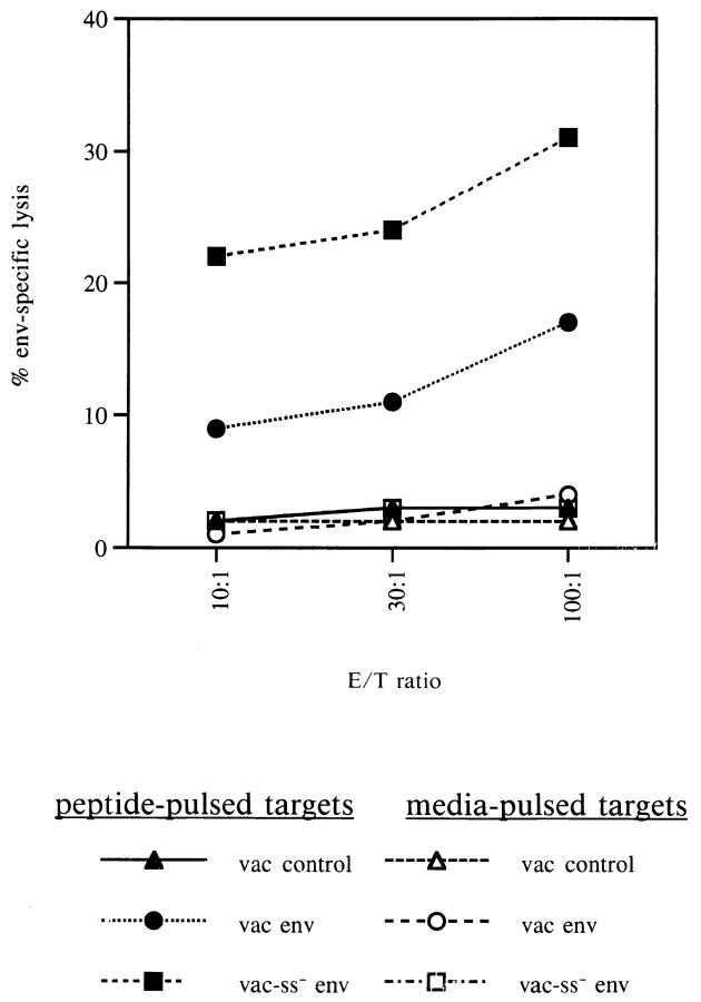
CD8+ cytotoxic T lymphocytes (CTLs) have the ability to recognize and eliminate virally infected cells before new virions are produced within that cell. Therefore, a rapid and vigorous CD8+ CTL response, induced by vaccination, can, in principle, prevent disseminated infection in vaccinated individuals who are exposed to the relevant virus. There has thus been interest in novel vaccine strategies that will enhance the induction of CD8+ CTLs. In this study, we have tested the hypothesis that targeting an antigen to undergo more efficient processing by the class I processing pathway will elicit a more vigorous CD8+ CTL response against that antigen. Targeting a type I transmembrane protein, the HIV-1 envelope (env) protein, for expression in the cytoplasm, rather than allowing its normal co-translational translocation into the endoplasmic reticulum, sensitized target cells expressing this mutant more rapidly for lysis by an env-specific CTL clone. Additionally, a greatly enhanced de novo env-specific CTL response was induced in vivo after immunization of mice with recombinant vaccinia vectors expressing the cytoplasmic env mutant. Similarly, targeting a cytoplasmic protein, HIV-1 nef, to undergo rapid cytoplasmic degradation induced a greatly enhanced de novo nef-specific CD8+ CTL response in vivo after immunization of mice with either recombinant vaccinia vectors or DNA expression plasmids expressing the degradation targeted nef mutant. The targeting of viral antigens for rapid cytoplasmic degradation represents a novel and highly effective vaccine strategy for the induction of enhanced de novo CTL responses in vivo.


Figures

Figure 1
Figure 1
Lysis of targets expressing wt or ss env protein by the envspecific CD8+ CTL clone A42.46. Autologous B-LCL were infected with the indicated vaccinia expression vectors 2 h before assay (diamonds) or were infected for 2 h and then incubated overnight at 37°C before assay (squares). Infected cells were then used as targets for the env-specific CD8+ CTL clone A42.46 in a standard 51Cr release assay at an E/T ratio of 10:1. One set of targets was infected in the presence of 0.1 mM cycloheximide (CHX, circles). The experiment was repeated three times with similar results. Data from a representative experiment are shown.
Figure 1
Figure 1
Lysis of targets expressing wt or ss env protein by the envspecific CD8+ CTL clone A42.46. Autologous B-LCL were infected with the indicated vaccinia expression vectors 2 h before assay (diamonds) or were infected for 2 h and then incubated overnight at 37°C before assay (squares). Infected cells were then used as targets for the env-specific CD8+ CTL clone A42.46 in a standard 51Cr release assay at an E/T ratio of 10:1. One set of targets was infected in the presence of 0.1 mM cycloheximide (CHX, circles). The experiment was repeated three times with similar results. Data from a representative experiment are shown.
Figure 2
Figure 2
Enhanced induction of env-specific CTLs by immunization with vac ss env. BALB/c mice were immunized with 107 PFU of vac control, vac-env, or vac-ss env. Splenocytes from immunized mice were stimulated for 5 d in IL-2 containing media in the presence of the H-2Ld– restricted env peptide (RGPGGRAFVTI). Stimulated splenocytes were then assayed for env-specific CTL activity against P815 cells pulsed with the H-2Ld–restricted env peptide or media alone. The data shown are representative of three separate experiments.
Figure 3
Figure 3
Precursor CTL (pCTL) frequency analysis. BALB/c mice were immunized with 107 PFU vac control, vac-env, or vac-ss env. After 21 d, splenocytes from immunized mice were cultured (48 wells/titration) in the presence of 105 naive splenocytes that had been pulsed with the H-2Ld–restricted env peptide for 2 h and then irradiated for 7 d. Culture media contained IL-2, T cell growth factor (from Con A–stimulated rat splenocytes), and methyl-α-d-mannopyranoside. Each well was then split into two equal parts and assayed for env-specific CTL activity against P815 targets pulsed with the H-2Ld–restricted env peptide or with media for 2 h. Positive wells were scored as >3 standard deviations above the percent specific lysis without effectors. The fraction of negative wells is plotted on a log scale versus cell number/well and from this line the pCTL frequency was determined (see Materials and Methods).
Figure 4
Figure 4
Enhanced stimulation of primary env-specific CTL response by immunization with vac-ss env. BALB/c mice were immunized with 107 PFU vac control, vac-env, or vac-ss env. After 6 d, splenocytes were isolated and assayed for direct CTL activity against peptide-pulsed (filled) or media-pulsed (open) P815 cells.
Figure 5
Figure 5
Pulse-chase analysis and immunoprecipitation of nef. P815 cells infected with vaccinia vectors expressing β-gal (lane 1), nef (lanes 2–5), UbMNef (lanes 6–9), and UbRNef (lanes 10–13) were pulse labeled with 35S-Met and 35S-Cys for 30 min and then chased with an excess of unlabeled Met and Cys for 0 min (lanes 1, 2, 6, 10), 15 min (lanes 3, 7, 11), 60 min (lanes 4, 8, 12) or 240 min (lanes 5, 9, 13), followed by extraction, immunoprecipitation, and electrophoretic analysis of nef and MHC class I expression (see Materials and Methods).
Figure 6
Figure 6
Stimulation of secondary nef-specific CTLs by UbMNef and UbRNef constructs. PBMCs were isolated from an HIV-1 seropositive donor and stimulated in the presence of IL-2 with titrating numbers of vVUbMNef (squares) or vVUbRNef (diamonds) infected, psoralen/UV–treated autologous B-LCL. Stimulated PBMCs were assayed for nef-specific CTL activity against vVnef infected (filled) or vac control infected (open) autologous B-LCL at an E/T ratio of 10.
Figure 6
Figure 6
Stimulation of secondary nef-specific CTLs by UbMNef and UbRNef constructs. PBMCs were isolated from an HIV-1 seropositive donor and stimulated in the presence of IL-2 with titrating numbers of vVUbMNef (squares) or vVUbRNef (diamonds) infected, psoralen/UV–treated autologous B-LCL. Stimulated PBMCs were assayed for nef-specific CTL activity against vVnef infected (filled) or vac control infected (open) autologous B-LCL at an E/T ratio of 10.
Figure 7
Figure 7
Enhanced induction of primary nef-specific CTLs by immunization with vVUbRNef. BALB/c mice were immunized with 107 PFU of vac control (open), vVUbMNef (stippled), or vVUbRNef (filled). Splenocytes from immunized mice were stimulated for 5 d in the presence of IL-2 with either CT26nef transfectants treated with mitomycin C or with CT26 cells which had been infected with vVnef and then treated with psoralen/UV light to inactivate vaccinia virus. The ratio of responders to stimulators was 50:1. Stimulated splenocytes were then assayed for nefspecific CTL activity against vVnef-infected CT26 cells or vac control– infected CT26 cells. The percentage nef-specific lysis reported is the percentage specific lysis of vVnef-infected targets − the percentage specific lysis of vac control–infected targets. The data shown are from a single experiment which is representative of three experiments conducted with similar results.
Figure 8
Figure 8
Enhanced induction of nef-specific CTLs by immunization with DNA expression vectors encoding UbRNef. BALB/c mice were immunized once with 4 × 50 μg pcDNA3 (open), pcDNA3nef (cross hatched), pcDNA3UbMNef (stippled), or pcDNA3UbRNef (filled). Splenocytes from immunized mice were stimulated with psoralen/UV–treated, vVnefinfected CT26 cells at a ratio of 50 responders/stimulator cell for 5 d in the presence of IL-2. Stimulated splenocytes were assayed for nef-specific CTL activity against CT26 cells infected with vVnef or with vac control or against P815 cells infected with vVnef or vac control at an E/T ratio of 10:1 or 100:1. The data shown are from one experiment. The experiment was repeated a total of four times, with similar results.

References

    1. Klavinskis LS, Tishon A, Oldstone MB. Efficiency and effectiveness of cloned virus-specific cytotoxic T lymphocytes in vivo. J Immunol. 1989;143:2013–2016. - PubMed
    1. Cannon MJ, Openshaw PJ, Askonas BA. Cytotoxic T cells clear virus but augment lung pathology in mice infected with respiratory syncytial virus. J Exp Med. 1988;168:1163–1168. - PMC - PubMed
    1. Riddell SR, Watanabe KS, Goodrich JM, Li CR, Agha ME, Greenberg PD. Restoration of viral immunity in immunodeficient humans by the adoptive transfer of T cell clones. Science (Wash DC) 1992;257:238–241. - PubMed
    1. Braciale TJ, Braciale VL. Viral antigen presentation and MHC assembly. Semin Immunol. 1992;4:81–84. - PubMed
    1. Yewdell JW, Bennink JR. Antigen processing: a critical factor in rational vaccine design. Semin Hematol. 1993;30:26–32. - PubMed

Publication types

MeSH terms