. 1997 Mar 18;94(6):2122-7.
doi: 10.1073/pnas.94.6.2122.

Removal of a cryptic intron and subcellular localization of green fluorescent protein are required to mark transgenic Arabidopsis plants brightly

Affiliations

Removal of a cryptic intron and subcellular localization of green fluorescent protein are required to mark transgenic Arabidopsis plants brightly

J Haseloff et al. Proc Natl Acad Sci U S A. .

Abstract

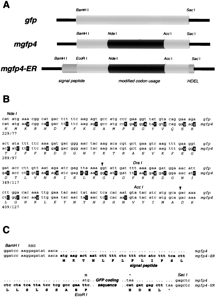
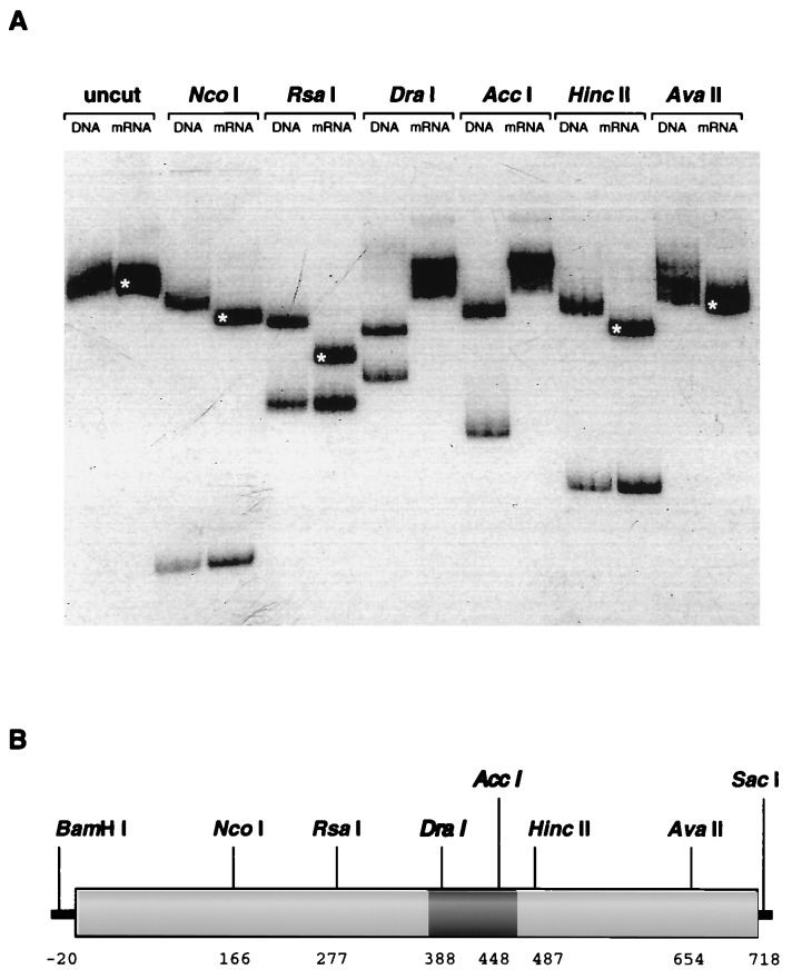
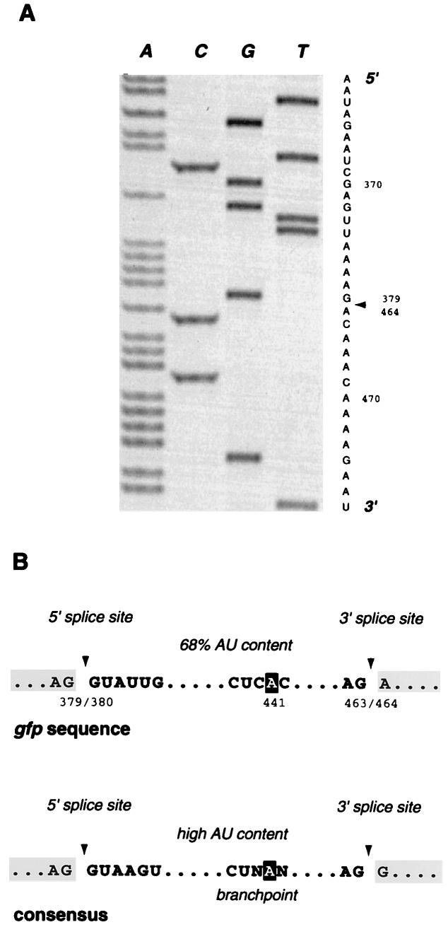
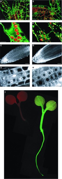
The green fluorescent protein (GFP) from the jellyfish Aequorea victoria is finding wide use as a genetic marker that can be directly visualized in the living cells of many heterologous organisms. We have sought to express GFP in the model plant Arabidopsis thaliana, but have found that proper expression of GFP is curtailed due to aberrant mRNA processing. An 84-nt cryptic intron is efficiently recognized and excised from transcripts of the GFP coding sequence. The cryptic intron contains sequences similar to those required for recognition of normal plant introns. We have modified the codon usage of the gfp gene to mutate the intron and to restore proper expression in Arabidopsis. GFP is mainly localized within the nucleoplasm and cytoplasm of transformed Arabidopsis cells and can give rise to high levels of fluorescence, but it proved difficult to efficiently regenerate transgenic plants from such highly fluorescent cells. However, when GFP is targeted to the endoplasmic reticulum, transformed cells regenerate routinely to give highly fluorescent plants. These modified forms of the gfp gene are useful for directly monitoring gene expression and protein localization and dynamics at high resolution, and as a simply scored genetic marker in living plants.


Figures

Figure 1
Figure 1
Modified gfp sequences. (A) Schematic diagrams showing gene expression cassettes that contain the wild-type sequence (gfp) (6), modified codon usage (mgfp4), and additional peptide targeting sequences (mgfp4-ER). The sequences that have altered codon usage are indicated in black, and targeting sequences are shown in dark grey. (B) Altered codon usage in mgfp4. The cryptic intron is shown underlined with the 5′ and 3′ splice sites arrowed. The upper sequence corresponds to that of gfp, and the lower nucleotide sequence is that of mgfp4. Mutated nucleotides are shown in reverse type, and restriction endonuclease sites are shadowed. The amino acid sequence is the same for each gene and is shown below. (C) Sequences flanking the GFP coding region. Both gfp and mgfp4 are flanked by restriction endonuclease sites for BamHI and SacI, a ribosome binding site (RBS) for bacterial expression, and the sequence AACA upstream of the start codon for improved plant translation. The mgfp4-ER gene cassette contains additional sequences shown in boldface type, which comprise a 5′ terminal signal peptide and 3′ HDEL sequence. An EcoRI site was used to link the signal peptide and coding sequences.
Figure 2
Figure 2
Aberrant posttranscriptional processing of gfp mRNA. (A) Restriction endonuclease digestion of PCR fragments derived from gfp DNA and mRNA sequences. Sequences corresponding to the integrated gfp gene and to mRNA transcripts were isolated and separately amplified using PCR techniques and incubated with various restriction endonucleases. The radiolabeled fragments were fractionated by electrophoresis in a 5% polyacrylamide gel, and are shown labeled with the source of the amplified sequences (DNA or mRNA) and the name of the restriction endonuclease used for digestion, or not (uncut). The mRNA-derived sequences appeared to lack sites for DraI and AccI and to contain deleted sequences. Fragments that are smaller than expected have been indicated with a white asterisk. (B) Schematic diagram of the gfp coding sequence. The positions of the restriction endonuclease cleavage sites that were used for analysis of PCR products are indicated, and these are numbered according to the coding region of gfp. The cleavage pattern of the amplified gfp mRNA sequence corresponds to a deletion of 80–90 nt, and this is indicated by a dark shaded region.
Figure 3
Figure 3
Sequence analysis of cloned gfp mRNAs. (A) Autoradiograph and sequence of the amplified gfp mRNA sequence. Nucleotides 380–463 are absent from the transcribed sequence, and the site of this 84-nt deletion is indicated by an arrowhead. (B) Comparison of gfp cryptic intron sequences with the conserved sequences that are found at the splice sites and branchpoints of plant introns (21, 22). Splice sites are indicated by arrowheads, and the nucleotide involved in branchpoint formation is shown in reverse type.
Figure 4
Figure 4
Confocal images of 35S-mgfp4 and 35S-mgfp4-ER transformed Arabidopsis plants. (AD) Images of 35S-mgfp4 transformed seedlings. (EH) Images of 35S-mgfp4-ER transformed seedlings. A and E show optical sections through the shoot apex of the seedlings, at the junction of cotyledons and hypocotyl (bars = 25 microns). Cells from the hypocotyl are shown in B (cortex) and F (epidermal; bars = 10 microns). Median longitudinal optical sections of root tips are shown in C and G (bars = 25 microns). Cells within the root tips are shown at higher magnification in D and H (bars = 10 microns). The distribution of the different forms of GFP is clear, with GFP accumulating within the nucleoplasm, but being excluded from the nucleolus (D), and with the ER-targeted form of GFP being excluded from the nucleus and forming a distinct perinuclear pattern. Similar partitioning of the two GFP forms can be seen in the shoot (A and E). (I) 5-day-old wild-type (Left) and 35S-mgfp4-ER transgenic (Right) seedlings were mounted in water on a Leitz DM-IL inverted fluorescence microscope and illuminated with long-wavelength UV light.

References

    1. Jefferson R A, Kavanagh T A, Bevan M W. EMBO J. 1987;6:3901–3907. - PMC - PubMed
    1. Heim R, Prasher D C, Tsien R Y. Proc Natl Acad Sci USA. 1994;91:12501–12504. - PMC - PubMed
    1. Cody C W, Prasher D C, Westler W M, Prendergast F G, Ward W W. Biochemistry. 1993;32:1212–1218. - PubMed
    1. Yang F, Moss L G, Phillips G N. Nat Biotechnol. 1996;14:1246–1220. - PubMed
    1. Ormo M, Cubitt A B, Kallio K, Gross L A, Tsien R Y, Remington S J. Science. 1996;273:1392–1395. - PubMed

Publication types

MeSH terms

Associated data