. 1997 Mar 18;94(6):2221-6.
doi: 10.1073/pnas.94.6.2221.

Regulation of E2F through ubiquitin-proteasome-dependent degradation: stabilization by the pRB tumor suppressor protein

Affiliations

Regulation of E2F through ubiquitin-proteasome-dependent degradation: stabilization by the pRB tumor suppressor protein

M R Campanero et al. Proc Natl Acad Sci U S A. .

Abstract

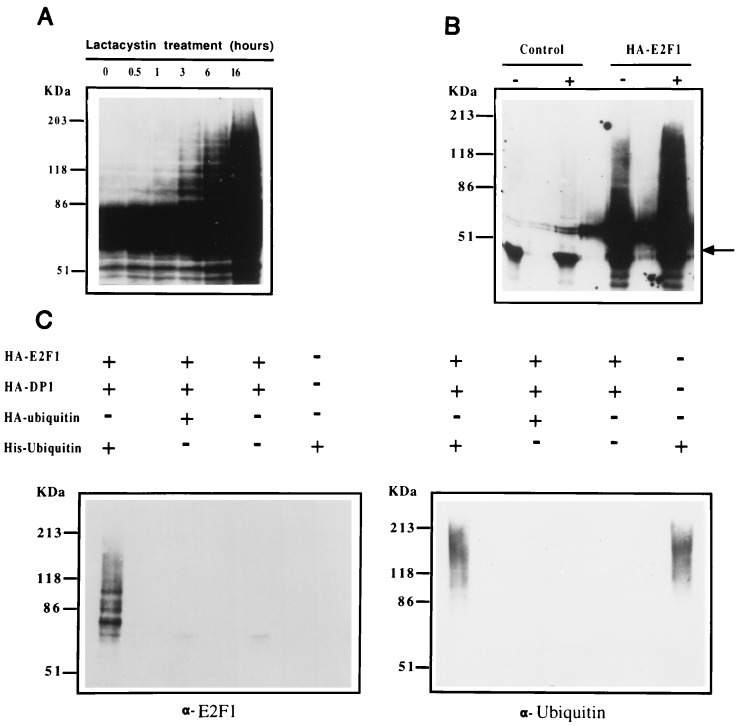
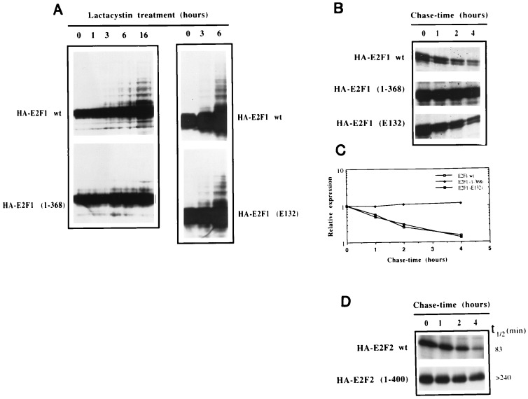
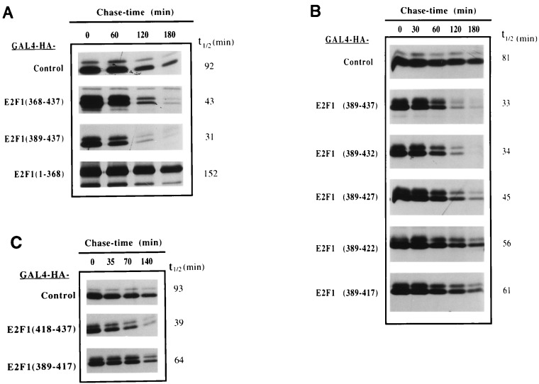
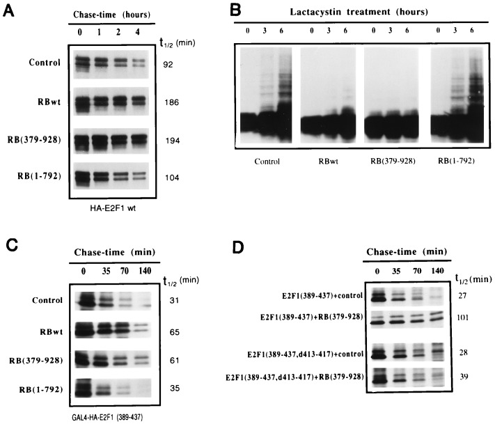
The E2F family of transcription factors plays a key role in regulating cell-cycle progression. Accordingly, E2F is itself tightly controlled by a series of transcriptional and posttranscriptional events. Here we provide evidence that E2FI protein levels are regulated by the ubiquitin-proteasome-dependent degradation pathway. An analysis of E2F1 mutants identified a conserved carboxyl-terminal region, which is required for eliciting ubiquitination and protein turnover. Fusion of this E2F1 carboxyl-terminal sequence to a heterologous protein, GAL4, resulted in destabilization of GAL4. Previous studies identified an overlapping region of E2F1 that facilitates complex formation with retinoblastoma tumor suppressor protein, pRB, and we found that pRB blocks ubiquitination and stabilizes E2F1. These results suggest a new mechanism for controlling the cell-cycle regulatory activity of E2F1.


Figures

Figure 1
Figure 1
E2F1 expression is modulated through the ubiquitin–proteasome pathway in vivo. (A) U2OS osteosarcoma cells were cotransfected with HA-E2F1 and HA-DP1 and treated for the indicated periods of time with 10 μM lactacystin. Aliquots from cell extracts were immunoblotted with the anti-E2F1 p98 polyclonal antibody (9). No signal was detected in the control experiment with mock-transfected cells (data not shown). (B) Control or HA-E2F1 expression plasmids were transfected into U2OS cells. Sixteen hours after transfection, cells were split and one-half were treated with 10 μM lactacystin for 3 hr. Extracts from both treated (+) and untreated (−) cells were prepared under denaturing conditions and immunoprecipitated with anti-HA 12CA5 mAb followed by immunoblotting with anti-E2F1 p98 antibody. The arrow points to a nonspecific band that likely corresponds to the immunoglobulin heavy chain. (C) U2OS cells were transfected with the indicated expression plasmids. Lactacystin (10 μM) was added for the last 3 hr of culture. Cells were lysed under denaturing conditions and extracts were purified over nickel-NTA beads. The purified material was assayed by immunoblot analysis employing anti-E2F1 p98 and anti-ubiquitin antibodies (Left and Right, respectively).
Figure 2
Figure 2
Deletion of the transactivation domain inhibits ubiquitination and stabilizes E2F1. (A) U2OS cells were transfected with the indicated plasmids together with HA-DP1 and treated with 10 μM lactacystin for the indicated periods of time. Aliquots from extracts were subjected to immunoblot analysis employing the anti-E2F1 mAb, SQ71. (B) U2OS cells were transfected with the indicated plasmids and split into four different plates the following day. Two days after transfection, cells were methionine-starved for 30 min and then pulsed for 60 min with [35S]methionine. Cells were then washed twice with PBS and chased for the indicated periods of time with culture medium. Extracts were prepared under denaturing conditions and immunoprecipitated with the anti-E2F1 SQ41 mAb. Samples were analyzed by SDS/PAGE and autoradiography. (C) The experiment shown in Fig. 2B was analyzed using a PhosphorImager to quantitate the amounts of wild-type and mutant derivatives of E2F1 that were immunoprecipitated at the different chase-time points. The abundance at each time point was calculated relative to the abundance at 0 hr. For each construct, the line graph was adjusted to an exponential model using a graphing program (Cricket graph). (D) Pulse-chase experiments were performed as described in Fig. 2B, except that the anti-HA 12CA5 mAb was used for immunoprecipitations. Half-life (t1/2) determinations were calculated as in Fig. 2C.
Figure 3
Figure 3
The E2F1 transactivation/pRb binding domain contains a degradation targeting sequence. U2OS cells were transfected with the indicated GAL4 or GAL4-E2F1 expression plasmids and split the day after into four or five plates. Pulse-chase experiments were carried out as described in Fig. 2. Extracts were prepared under nondenaturing conditions and immunoprecipitated with the anti-HA 12CA5 mAb. Samples were analyzed by SDS/PAGE and half-life (t1/2) determination was performed as described in Fig. 2.
Figure 4
Figure 4
The tumor suppressor protein pRb blocks ubiquitination and increases the half-life of E2F1. (A) The indicated pRb expression plasmids were cotransfected with HA-E2F1 and HA-DP1 into U2OS cells. The following day, cells were split into four plates for pulse-chase analysis. Extracts were prepared under denaturing conditions and immunoprecipitated with the SQ41 anti-E2F1 mAb. Samples were analyzed by SDS/PAGE and autoradiography. Half-lives (t1/2) were calculated as indicated in Fig. 2. (B) Expression plasmids for HA-E2F1, together with HA-DP1 and the indicated pRB constructs, were cotransfected into U2OS cells. Lactacystin (10 μM) was added for the indicated periods of time. Aliquots from cell extracts were immunoblotted with anti-E2F1 SQ71 mAb. (C) The indicated pRb expression plasmids were cotransfected with GAL4-HA-E2F1(389–437) into U2OS cells. The following day, cells were split into four plates for pulse-chase analysis. Extracts were prepared under nondenaturing conditions and immunoprecipitated with the 12CA5 anti-HA mAb. (D) GAL4-E2F1(389–437) or the pRb binding mutant GAL4-E2F1(389–437,d413–417) plasmids were cotransfected with a control or a pRb(379–928) expression vector into U2OS cells. Cells were cotransfected with non-HA-tagged pRB expression vectors so that coimmunoprecipitation/interaction studies could be performed.

References

    1. Nevins J R. Science. 1992;258:424–429. - PubMed
    1. Weintraub S J, Prater C A, Dean D C. Nature (London) 1992;358:259–261. - PubMed
    1. Weintraub S J, Chow K N B, Luo R X, Zhang S H, He S, Dean D C. Nature (London) 1995;375:812–815. - PubMed
    1. Sellers W R, Rodgers J W, Kaelin W G. Proc Natl Acad Sci USA. 1995;92:11544–11548. - PMC - PubMed
    1. Weinberg R A. Cell. 1995;81:323–330. - PubMed

Publication types

MeSH terms

LinkOut - more resources