. 1997 Mar 18;94(6):2295-300.
doi: 10.1073/pnas.94.6.2295.

Stat5 is a physiological substrate of the insulin receptor

Affiliations

Stat5 is a physiological substrate of the insulin receptor

J Chen et al. Proc Natl Acad Sci U S A. .

Abstract

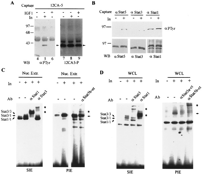
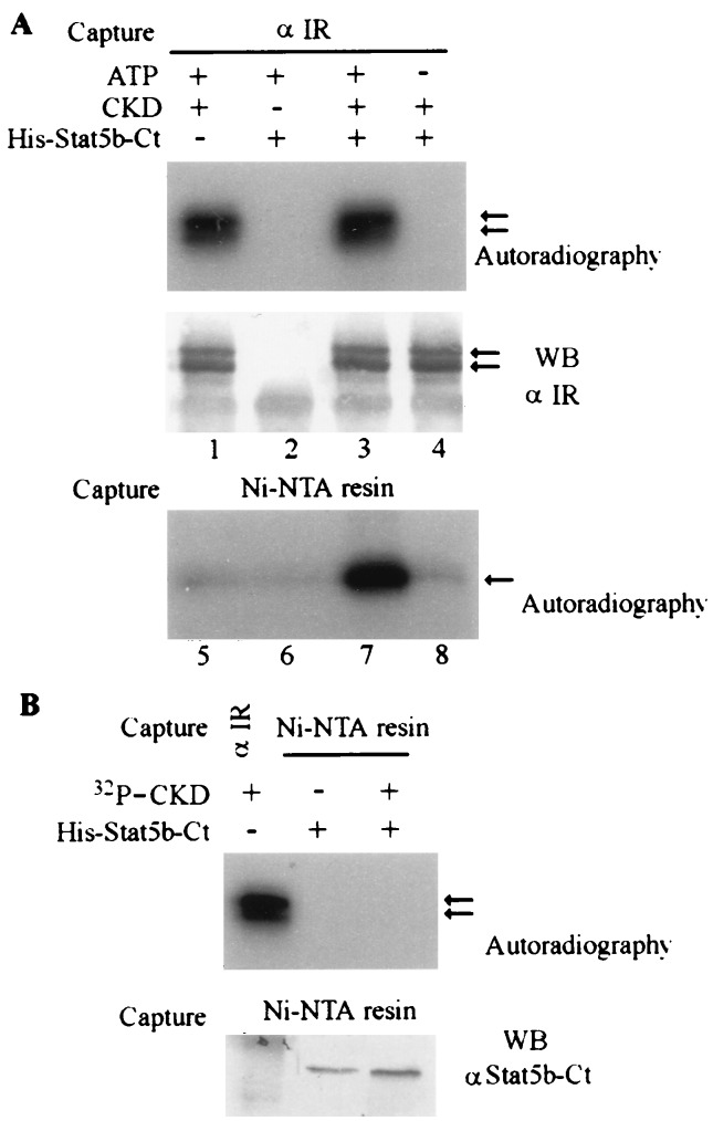
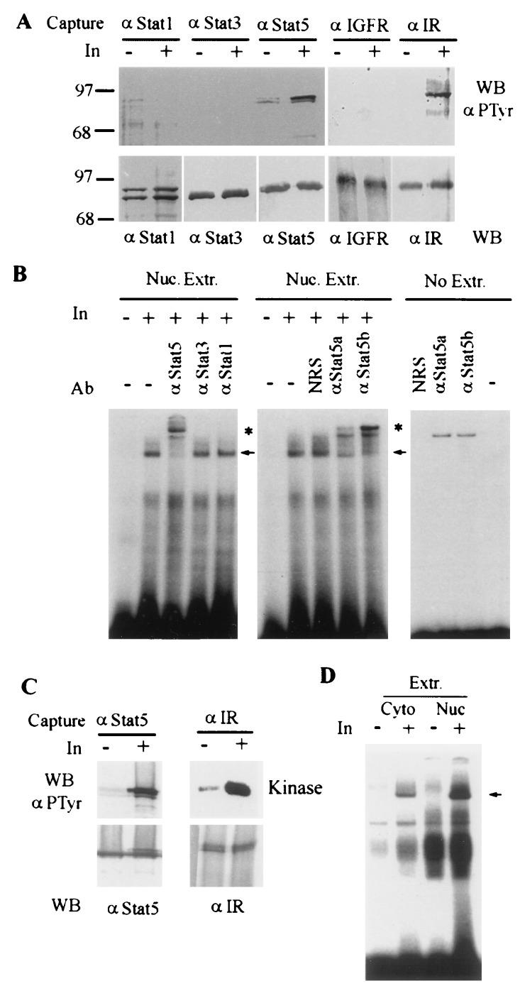
Using the cytoplasmic domain of the insulin receptor (IR) in a yeast two-hybrid screen, we identified a cDNA clone encoding the C-terminal 308 amino acids of human Stat5b (Stat5b-Ct). Stat5b-Ct is tyrosine phosphorylated by purified IR kinase domain in vitro. Insulin stimulates tyrosine phosphorylation of overexpressed Stat5b-Ct and endogenous Stat5 in cells overexpressing IR. Stat5 may be a direct target of the IR and, as a member of the Stat family of transcription factors, may play a role in the regulation of gene transcription by insulin. In support of this hypothesis, perfusion of mouse liver with insulin promotes rapid tyrosine phosphorylation of Stat5 and activation of Stat5 DNA binding. Moreover, refeeding of fasted mice leads to rapid tyrosine phosphorylation and stimulation of enhanced DNA-binding activity of Stat5 extracted from liver, skeletal muscle, and adipose tissues. Taken together, our data strongly suggest that IR interacts with and phosphorylates Stat5 in vitro and in tissues physiologically sensitive to insulin.


Figures

Figure 3
Figure 3
Stat5b-Ct and endogenous Stat5b are tyrosine phosphorylated in response to insulin stimulation in vivo. (A) Tyrosine phosphorylation of Stat5b-Ct in COS cells. Five micrograms each of pRcCMV-Stat5b-Ct and pECE-IR were cotransfected into COS1 cells. After 48 h, the cells were starved for 2 h in serum-free medium and then stimulated with either insulin (50 nM) or IGF1 (20 ng/ml) for 15 min. HA-Stat5b-Ct was immunoprecipitated with 12CA5 antibody, and the immunoprecipitate was divided into two aliquots, one for Western blotting with RC20 (Left) and the other with horseradish peroxidase-12CA5 (Right). Arrows indicate the positions of Stat5b-Ct. (B) Tyrosine phosphorylation of Stat3 and Stat5 in CHO-IR cells. CHO-IR cells were starved in serum-free medium for 4 h in the presence of 200 mM Na3VO4 and then stimulated with insulin (50 nM) for 15 min. One microgram of protein each from the cell lysates was immunoprecipitated with anti-Stat1, Stat3, or Stat5 antibodies. Each immunoprecipitate was divided into two equal parts for detection of Stat tyrosine phosphorylation (Upper) and Stat protein (Lower). (C) Insulin activates Stat3 and Stat5 DNA binding in CHO-IR cells. CHO-IR cells were treated as in B, and nuclear extracts were prepared. Nuclear extracts (20 μg) were subjected to electrophoretic mobility-shift assay (EMSA) analysis with the m67 SIE (Left) or β-casein PIE (Right) probes. Anti-Stat1 and Stat3 antibodies and an antibody against the N-terminal portion of Stat5b (Stat5b-nt) (recognizes both murine Stat5a and Stat5b) were used for the supershift assays. (D) Insulin activates Stat1, Stat3, Stat5a, and Stat5b DNA-binding in HIR3.5 cells. Cells were starved in DMEM with 0.1% BSA for 4 h and then treated with 200 μM vanadate for 40 min, when buffer or insulin (50 nM) was added for the last 15 min. Whole cell lysates were prepared and subjected to gel-mobility shift analysis with the M67 SIE (Left) and β-casein PIE (Right) probes. Antibodies against Stat1, Stat3, Stat5a-Ct, and Stat5b-Ct were used for supershift. Arrows indicate the specific insulin-inducible complexes; asterisks indicate supershifted Stat-containing complexes.
Figure 1
Figure 1
Schematic representation of plasmids used in the two-hybrid system pLexA-IR-S and pLexA-IR-L are the bait fusion plasmids containing the short or long fragment of the insulin receptor β subunit, respectively. pJG4–5-cDNA is the cDNA library derived from HeLa cells mRNAs. pSH18–34 is the reporter plasmid. EC, extracellular; TM, transmembrane domain; PTK, protein tyrosine kinase; MCS, multiple cloning sites; NLS, nuclear localization signal; AD, activation domain; HA, hemagglutinin tag sequence; LexA BS, LexA binding site.
Figure 2
Figure 2
In vitro phosphorylation assay of His-Stat5b-Ct. (A) His-Stat5b-Ct can be phosphorylated by CKD in vitro. First 0.3 μg of CKD and 0.75 μg of His-Stat5b-Ct proteins were incubated in 50 μl of kinase buffer (10 mM MnCl2/50 mM Tris·HCl, pH 8.0). [γ-32P]ATP (10 μCi) was added to start the reaction. At the end of reaction, 0.5 ml of RIPA buffer was added, and CKD protein was first immunoprecipitated by anti-IR. (Upper) Kinase assay. (Lower) Amount of CKD protein immunoprecipitated and detected by Western blotting with anti-IR antibody. His-Stat5b-Ct protein subsequently was captured from the supernatant by Ni-NTA-resin, washed with 6 M guanidine hydrochloride, followed by water, and then eluted with Laemmli buffer. The phosphorylated His-Stat5b-Ct was detected by autoradiography. (B) His-Stat5b-Ct does not coprecipitate with CKD in vitro. His-Stat5b-Ct was incubated with preautophosphorylated CKD in RIPA buffer and precipitated with Ni-NTA-resin as above. (Upper) Autoradiography. The left lane is the positive control for 32P-CKD precipitated with anti-IR Ab. (Lower) His-Stat5b-Ct protein captured by Ni-NTA-resin and detected by Western blotting with anti-Stat5b-Ct Ab.
Figure 4
Figure 4
Insulin stimulates Stat5 tyrosine phosphorylation and DNA binding activity in perfused liver. (A) Stat5 is tyrosine phosphorylated in mouse liver perfused with insulin. The livers were perfused with 10 ml Hanks solution containing either 1 mM Na3VO4 or 1 mM Na3VO4 plus insulin (50 nM). After 10 min, the liver lysates were prepared. Three milligrams of protein of each lysate was immunoprecipitated with antibodies against Stat1, Stat3, Stat5, IR, or IGF1 receptor and then divided into two parts, one part for Western blotting to detect tyrosine phosphorylation (Upper) and the other for assessing protein amount (Lower). (B) Insulin stimulates Stat5 DNA-binding activity. Nuclear proteins were extracted from the perfused mouse liver and subjected to EMSA using a 32P-labeled double-stranded oligonucleotide probe that contained a MGF/Stat5 binding site from the β-casein promoter (PIE) (29). The Stat-specific antibodies were used for supershift assay. The nonspecific supershift was determined in the absence of nuclear extract. (C) Stat5 is tyrosine phosphorylated in mouse liver perfused with insulin in the absence of sodium vanadate. Conditions were the same as in A except sodium vanadate was not added in the perfusion buffer, and 6 mg of total protein of each lysate was used for analysis. (D) Insulin stimulates Stat5 DNA binding in the absence of sodium vanadate. Frozen pieces of the mouse livers perfused as described in C were thawed and cytoplasmic and nuclear extracts were prepared. Stat5 DNA-binding activity in these extracts (30 μg) was assessed similarly using PIE probe. Arrows indicate the specific insulin-inducible complexes; asterisks indicate supershifted Stat5-containing complexes.
Figure 5
Figure 5
Refeeding of the fasted mice leads to Stat5 tyrosine phosphorylation and activation in insulin target tissues. (A) Tyrosine phosphorylation of Stat5 in liver, muscle, and adipose tissues of refed mice. Mice were starved for 3 days, allowed to feed for 80 min, and then sacrificed. Whole cell lysates were prepared from the indicated tissues. Three milligrams each of the total protein lysates was used for immunoprecipitation with anti-Stat5. A second immunoprecipitation from the supernatant was then performed with anti-IR antibody. Anti-Stat5 immunoprecipitates were divided into two parts: one part for PTyr Western blotting (Top; faint signal of Stat5 tyrosine phosphorylation in the heart of refed mice in the original filter did not reproduce well), and the other part for anti-Stat5 blotting (Upper Middle). The complexes captured by anti-IR was assayed in vitro for kinase activity (Lower Middle). The same filter was used for detecting the IR amount by Western blotting with anti-IR antibody (Bottom). (B) Stat5 DNA binding activity is induced by refeeding. Cytoplasmic and nuclear extracts from the livers of fasted and refed mice were subjected to EMSA analysis with the PIE probe (29). Arrow indicates the refeeding-induced complex.

Similar articles

Cited by

References

    1. Draznin B, Melmed S, Leroith D, editors. Insulin Action, Molecular and Cellular Biology of Diabetes Mellitus. II. New York: Liss; 1989.
    1. Ebina Y, Ellis L, Jarnagin K, Edery M, Graf L, Clauster E, Ou J H, Masiarz F, Kan Y W, Goldfine I D, Roth R A, Rutter W J. Cell. 1989;40:747–758. - PubMed
    1. Chou C K, Dull T J, Russel D S, Gherzi R, Lebwohl D, Ullrich A, Rosen O M. J Biol Chem. 1987;262:1842–1847. - PubMed
    1. Ebina Y, Araki E, Taira M, Shimada F, Mori M, Craik C S, Siddle K, Pierce S B, Roth R A, Rutter W J. Proc Natl Acad Sci USA. 1987;84:704–708. - PMC - PubMed
    1. Cheatham B, Kahn C R. Endocrine Review. 1995;16:117–142. - PubMed

Publication types

MeSH terms

LinkOut - more resources