. 1997 Mar 18;94(6):2350-5.
doi: 10.1073/pnas.94.6.2350.

Dissecting the thrombopoietin receptor: functional elements of the Mpl cytoplasmic domain

Affiliations

Dissecting the thrombopoietin receptor: functional elements of the Mpl cytoplasmic domain

J G Drachman et al. Proc Natl Acad Sci U S A. .

Abstract

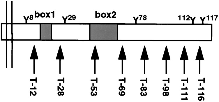
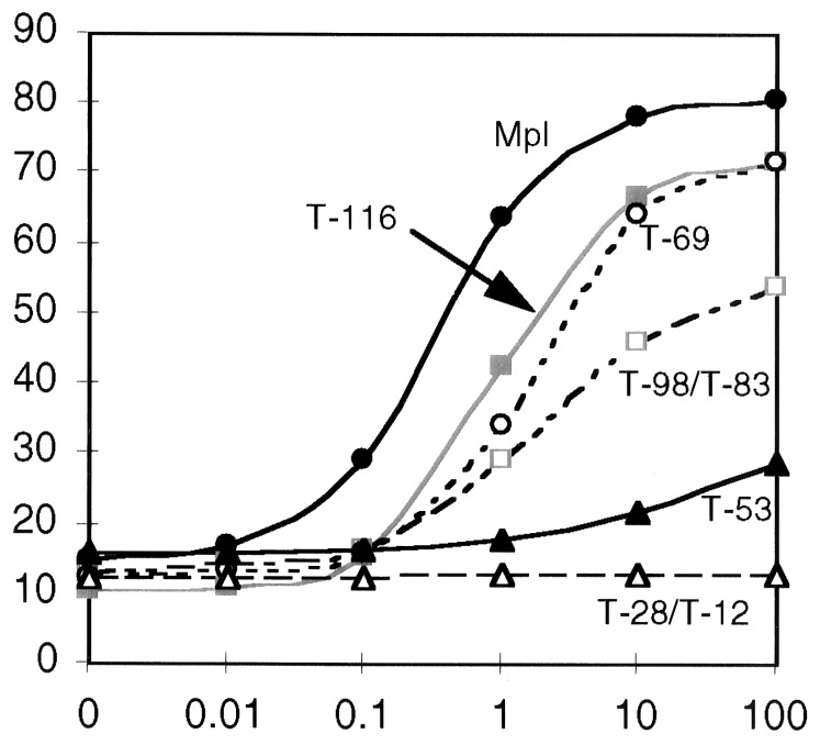
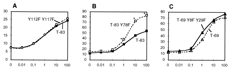
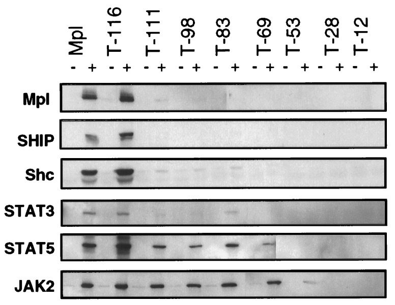
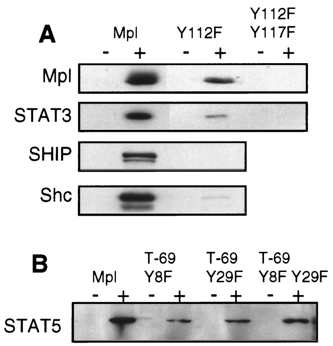
Thrombopoietin (TPO) acts through its receptor, Mpl, to stimulate the proliferation and maturation of megakaryocytes and their progenitors. The Mpl cytoplasmic domain controls this process through assembly of an active signaling complex using various receptor docking sites. In this report, eight carboxyl truncations of the 121-aa murine Mpl cytoplasmic domain were tested for the ability to support growth of a cytokine-dependent cell line (Ba/F3) and for their capacity to induce TPO-stimulated tyrosine phosphorylation of specific signaling proteins. Point mutations of the five tyrosine residues in the cytoplasmic domain of the receptor were subsequently used to confirm our conclusions. From these studies we demonstrate that: (i) TPO-induced proliferation is moderately reduced by truncation of as many as 53 C-terminal amino acids of Mpl, including the sites of receptor tyrosine phosphorylation; (ii) truncation/mutation of residues 69-83 of the Mpl cytoplasmic domain enhances proliferative signaling, perhaps mediated by a decrease in receptor-driven cellular differentiation; (iii) Mpl can be phosphorylated at either Y112 or Y117 but not at the three proximal cytoplasmic tyrosine residues (Y8, Y29, and Y78); (iv) Y112 of Mpl is necessary for tyrosine phosphorylation of Shc and Shc-associated p145 (SHIP); and (v) unlike STAT3, STAT5 is partially phosphorylated in the absence of any tyrosine residues in the Mpl cytoplasmic domain. These studies identify subdomains of Mpl necessary for activation of several critical signaling pathways and point to two potentially novel mechanisms of TPO-induced signal transduction, an indirect pathway to STAT5 activation and a differentiation domain that acts by limiting proliferation.


Figures

Figure 1
Figure 1
Cytoplasmic domain of Mpl is represented with cell membrane on the left and C terminus on the right. The five cytoplasmic tyrosine residues (Y), putative box1 and box2 motifs, and the location of the receptor truncations (arrows) are indicated. The truncations indicate the number of cytoplasmic residues that remain.
Figure 2
Figure 2
MTT assay of the Mpl truncations. The abcissa shows TPO concentration (0–100 ng/ml), and ordinate indicates percentage of maximum IL-3-induced proliferation. For clarity, curves that are essentially overlapping are represented by a single line. Proliferation curves are shown for Ba/F3 cells bearing the following receptors: full-length Mpl (•), T-116 (▪), T-98 and T-83 (□), T-69 (○), T-53 (▴), and T-28 and T-12 (▵). The data represent the mean values of four separate experiments.
Figure 3
Figure 3
Effect of tyrosine substitution on proliferation. For each assay, the abcissa shows TPO concentration (ng/ml), and the ordinate shows the percentage of maximal IL-3-induced proliferation. (A) MTT assay comparing T-83 (•) and full-length Mpl with two point mutations, Y112F Y117F (○). The curves represent combined results of two experiments. (B) Comparison of T-83 (▪) and T-83 Y78F (□). Combined results of three experiments are depicted; asterisks indicate significant difference between curves (P < 10−6). (C) Comparison of T-69 (▵) and T-69 Y8F Y29F (▴). Combined results of three experiments are shown. (Data in A was obtained in different experiments from those in panels B and C).
Figure 4
Figure 4
TPO-induced tyrosine phosphorylation of Mpl, SHIP, Shc, STAT3, STAT5, and JAK2. The proteins indicated on the left were immunoprecipitated from cellular lysates of Ba/F3 cells bearing either the full-length receptor (Mpl) or one of the truncations. Paired lanes indicate samples that had been unstimulated (−) or TPO-exposed (+). The Western blots were probed with a phosphotyrosine-specific antibody. Blots were stripped and reprobed to ensure equal protein in each lane (data not shown).
Figure 5
Figure 5
(A) Effect of Y112F or Y112F Y117F substitution on tyrosine phosphorylation. Lysates from cells expressing either the wild-type receptor (Mpl), the Y112F substitution mutant, or the Y112F Y117F double mutation were used to compare TPO-induced tyrosine phosphorylation of Mpl, STAT3, SHIP, and Shc. (B) TPO-induced STAT5 phosphorylation was compared in cells containing the full-length receptor (Mpl) or a truncated receptor in which one or both of the cytoplasmic tyrosine residues had been replaced by phenylalanine. Paired lanes indicate samples that had been unstimulated (−) or TPO-exposed (+). The Western blot was probed with a phosphotyrosine-specific antibody. Blots were stripped and reprobed to ensure equal protein in each lane (data not shown).

References

    1. Kaushansky K, Lok S, Holly R D, Broudy V C, Lin N, et al. Nature (London) 1994;369:568–571. - PubMed
    1. Kaushansky K, Broudy V, Lin N, Jorgensen M, McCarty J, Fox N, Zucker-Franklin D, Lofton-Day C. Proc Natl Acad Sci USA. 1995;92:3234–3238. - PMC - PubMed
    1. Alexander W S, Metcalf D, Dunn A R. EMBO J. 1995;14:5569–5578. - PMC - PubMed
    1. Broudy V C, Lin N L, Fox N, Taga T, Saito M, Kaushansky K. Blood. 1996;88:2026–2032. - PubMed
    1. Morella K K, Bruno E, Kumaki S, Lai C F, Fu J, Wang H M, Murray L, Hoffman R, Timour M, Bénit L, Gisselbrecht S, Zhvans H, Wojchowski D M, Bauman H, Gearing D P. Blood. 1995;86:557–571. - PubMed

Publication types

MeSH terms