. 1997 Mar 18;94(6):2392-7.
doi: 10.1073/pnas.94.6.2392.

Function and regulation of heat shock factor 2 during mouse embryogenesis

Affiliations

Function and regulation of heat shock factor 2 during mouse embryogenesis

M Rallu et al. Proc Natl Acad Sci U S A. .

Abstract

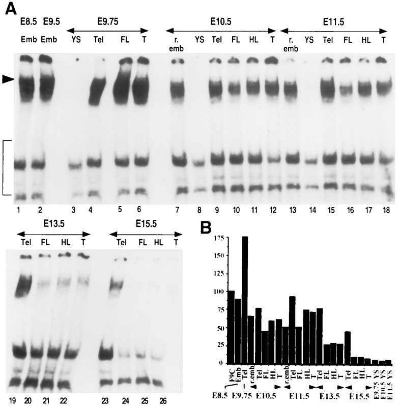
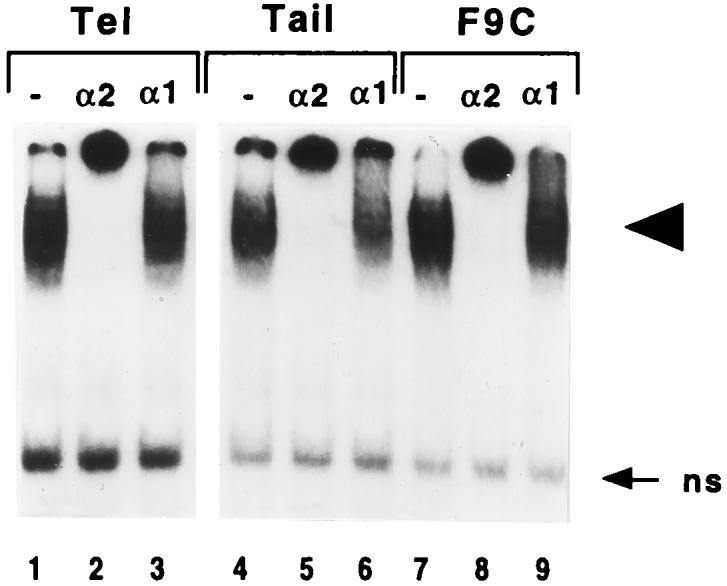
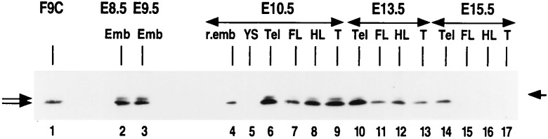
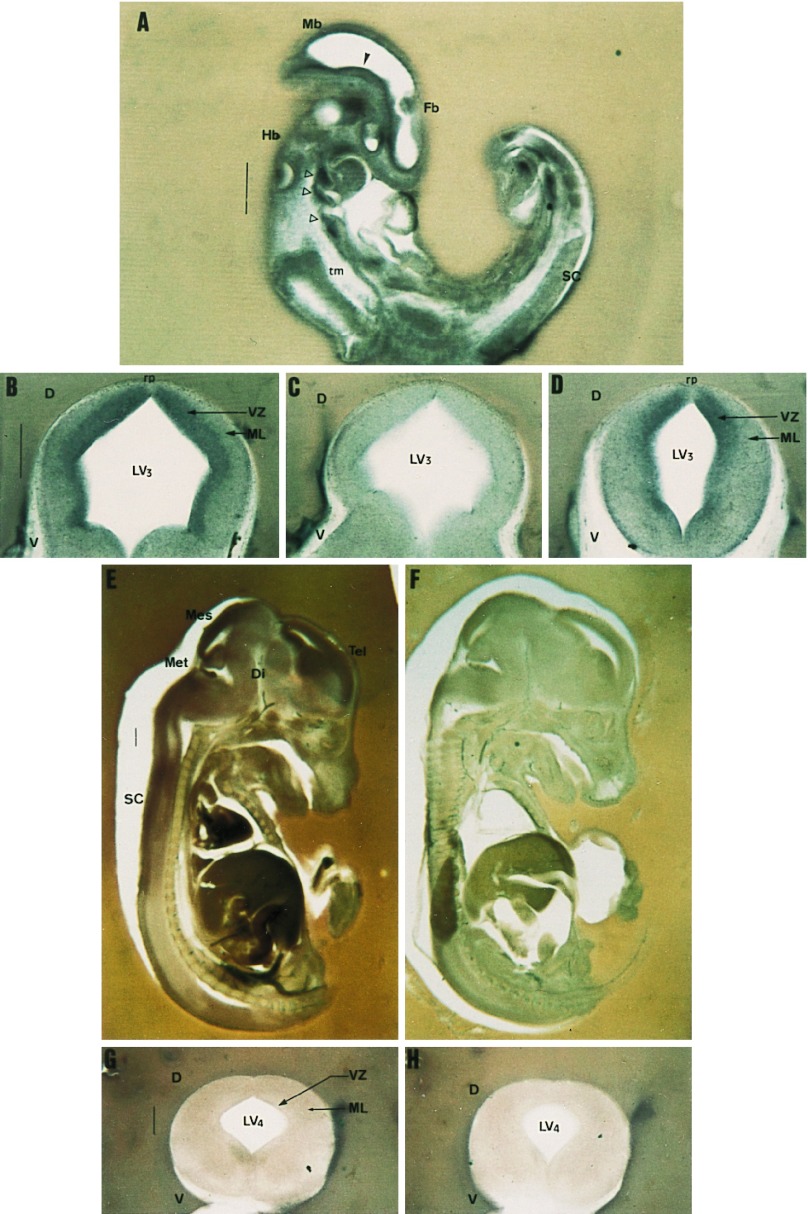
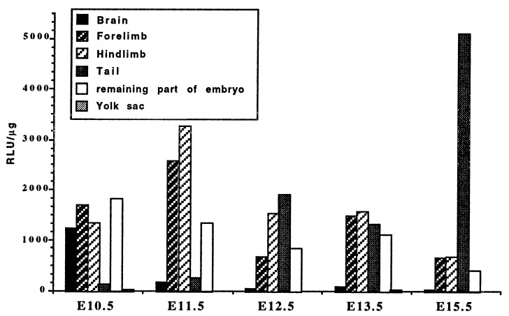
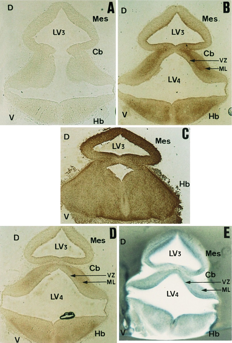
The spontaneous expression of heat shock genes during development is well documented in many animal species, but the mechanisms responsible for this developmental regulation are only poorly understood. In vertebrates, additional heat shock transcription factors, distinct from the heat shock factor 1 (HSF1) involved in the stress response, were suggested to be involved in this developmental control. In particular, the mouse HSF2 has been found to be active in testis and during preimplantation development. However, the role of HSF2 and its mechanism of activation have remained elusive due to the paucity of data on its expression during development. In this study, we have examined HSF2 expression during the postimplantation phase of mouse development. Our data show a developmental regulation of HSF2, which is expressed at least until 15.5 days of embryogenesis. It becomes restricted to the central nervous system during the second half of gestation. It is expressed in the ventricular layer of the neural tube which contains mitotically active cells but not in postmitotic neurons. Parallel results were obtained for mRNA, protein, and activity levels, demonstrating that the main level of control was transcriptional. The detailed analysis of the activity of a luciferase reporter gene under the control of the hsp70.1 promoter, as well as the description of the protein expression patterns of the major heat shock proteins in the central nervous system, show that HSF2 and heat shock protein expression domains do not coincide. This result suggests that HFS2 might be involved in other regulatory developmental pathways and paves the way to new functional approaches.


Figures

Figure 1
Figure 1
Detection of constitutive HSE-binding activity by gel shift assay in postimplantation embryos. Emb, entire embryo at E8.5 or E9.5; r.emb, remaining undissected part of the embryo; YS, yolk sac; Tel, telencephalic vesicles; FL, forelimbs; HL, hindlimbs; T, tail bud. (A) Extracts (27 μg) from dissected structures were incubated with 32P-labeled double-stranded HSE2 oligonucleotide for gel shift assay analysis. Arrowhead points to the specific shifted complexes. Nonspecific complexes are bracketed. Free DNA is not shown. (B) Results of PhosphorImager quantification of the specific shifted complexes. The signal intensities of embryo extracts were reported as the percentage of the density value in F9 control cell extracts.
Figure 2
Figure 2
Effect of polyclonal antibodies on specific HSE complexes formed with extracts from postimplantation embryos. Extracts (27 μg) from the telencephalic or tail parts of E11.5 embryos or from F9 control cells (F9C) were preincubated with polyclonal antibodies raised against HSF1 (α1) or HSF2 (α2) or without antibody (−), before incubation with the 32P-labeled double-stranded HSE2 oligonucleotide and gel shift assay analysis. Arrowhead points to the specific complexes, and arrow points to nonspecific complexes (ns).
Figure 3
Figure 3
Western blot analysis of HSF2 distribution in postimplantation embryos. F9C, extracts from F9 control cells. Left arrows indicate both HSF2 splicing isoforms; the right arrow indicates the 80-kDa molecular mass marker.
Figure 4
Figure 4
HSF2 ISH during postimplantation development. Sections 100–250 μm thick of E9.5 (A), E12.5 (BD), and E15.5 (EH) embryos were hybridized with digoxygenin-labeled antisense or sense (C, F, and H) HSF2 probes. (A) Parasagittal section of E9.5 embryo; HSF2 is expressed throughout the embryo except in the trunk mesenchyme. Arrowheads point to structures where HSF2 is highly expressed (see text). (BD) Coronal sections of E12.5 dissected neural tube at the level of the mesencephalon (B and C) or spinal cord (D). The expression domain maps to proliferative regions of the neural tube. Note that the roofplate region (rp) is not stained. (E and F) Sagittal sections of E15.5 embryos display markedly decayed HSF2 expression. Weak staining is only detectable in the different developing encephalic vesicles and spinal cord. (G and H) Coronal sections of E15.5 neural tubes (at the level of spinal cord) show diffuse low labeling. D, dorsal; Di, diencephalon; Fb, forebrain; Hb, hindbrain; LV3, mesencephalic vesicle; LV4, fourth ventricle; Mb, midbrain; Mes, mesencephalon; Met, metencephalon; ML, mantle layer; rp, roofplate; SC, spinal cord; Tel, telencephalon; tm, trunk mesenchyme; V, ventral; VZ, ventricular zone. (Bar = 500 μm.)
Figure 5
Figure 5
Luciferase activity in tissues of HSP70.1-luciferase transgenic embryos. Each value is the mean of the measurements performed with three embryos. The variations between embryos are <20%.
Figure 6
Figure 6
Comparative distribution profiles of the major HSPs and HSF2 gene expression within the CNS of E12.5 embryos. (AE) Immunological detection of HSPs on coronal cryostat sections of E12.5 dissected neural tubes at level of midbrain. Sections were incubated with anti-HSP90α (A), anti-HSC70 (B), anti-HSP90β (C), or anti-HSP25 (D). (E) ISH with digoxygenin-labeled HSF2 antisense riboprobe on a 200-μm-thick coronal section, performed at the level of midbrain Cb, cerebellum; other abbreviations are identical to those in Fig. 4.

References

    1. Pauli D, Tissières A. In: Developmental Expression of the Heat Shock Genes in Drosophila melanogaster. Morimoto R I, Tissières A, Georgopoulos C, editors. Plainview, NY: Cold Spring Harbor Lab. Press; 1990. pp. 361–378.
    1. Nakai A, Morimoto R I. Mol Cell Biol. 1993;13:1983–1997. - PMC - PubMed
    1. Rabindran S K, Giorgi G, Clos J, Wu C. Proc Natl Acad Sci USA. 1991;88:6906–6910. - PMC - PubMed
    1. Sarge K D, Zimarino V, Holm K, Wu C, Morimoto R I. Genes Dev. 1991;5:1902–1911. - PubMed
    1. Schuetz T J, Gallo G J, Sheldon L, Tempst P, Kingston R E. Proc Natl Acad Sci USA. 1991;88:6911–6915. - PMC - PubMed

Publication types

MeSH terms

LinkOut - more resources