. 1997 Mar 18;94(6):2489-94.
doi: 10.1073/pnas.94.6.2489.

Genetically engineered superantigens as tolerable antitumor agents

Affiliations

Genetically engineered superantigens as tolerable antitumor agents

J Hansson et al. Proc Natl Acad Sci U S A. .

Abstract

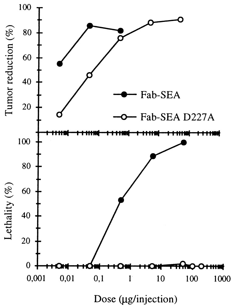
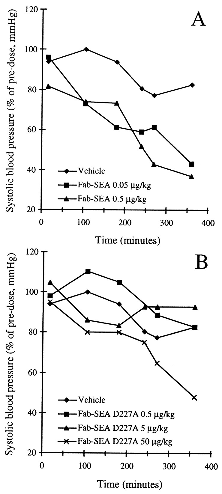
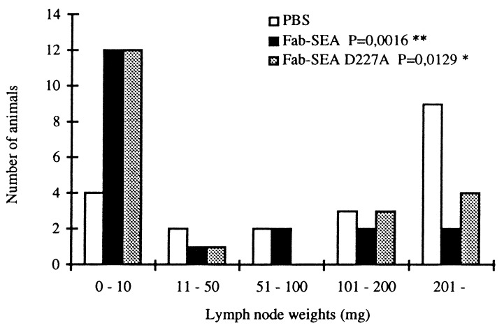
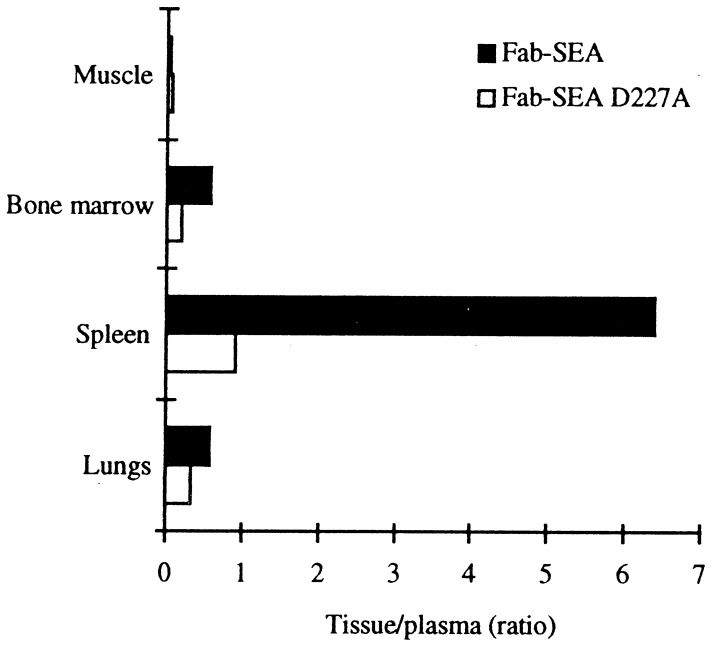
Superantigens (SAg) are a family of bacterial and viral proteins with strong immunostimulatory properties. SAg bound to major histocompatibility complex (MHC) class II molecules activate a high frequency of T cells and represent the most potent known activators of T cells to date. To explore the use of SAg for T cell-based tumor therapy we have created a tumor-reactive SAg by engineering a fusion protein composed of a tumor-reactive mAb (C215Fab) and the bacterial SAg staphylococcal enterotoxin A (SEA). A point mutation D227A was introduced at the major MHC class II binding site in SEA to reduce systemic toxicity. Treatment of tumor bearing mice with the Fab-SEA D227A fusion protein resulted in profound antitumor effects with a markedly reduced toxicity as compared with the wild-type Fab-SEA fusion protein. The reduced toxicity was probably due to a weak distribution of the SEA D227A fusion protein in tissues with a high MHC class II expression and low systemic cytokine levels as exhibited in mice and rabbits. The data presented demonstrate the efficacy of immunoconjugates containing a mutated SAg in directing a T cell attack against tumor cells with minimal systemic immune activation.


Figures

Figure 1
Figure 1
Tumor therapy and toxicity with C215Fab–SEA (Fab–SEA) and mutated C215Fab–SEA D227A (Fab–SEA D227A) in TCR Vβ3 TG mice. Reduction of pulmonary B16-C215 metastases are recorded after three injections of Fab–SEA proteins on a daily basis starting 1 day after tumor injection. Toxicity was noted after the third injection of Fab–SEA or Fab–SEA D227A in tumor-bearing TCR Vβ3 TG mice. Presented data represents the mean of three to four different experiments with six to eight animals in each experimental group.
Figure 2
Figure 2
Drop in systolic blood pressure after injection of Fab–SEA (A) or Fab–SEA D227A (B) in anesthesized New Zealand White rabbits. The change in blood pressure is presented as percent of stable pre-dose pressure of each individual animal.
Figure 3
Figure 3
Tumor therapy of established lymph node B16-C215 tumors in TCR Vβ3 TG mice. Lymph nodes were microinjected with B16-C215 tumor cells and screened for tumor growth after 5 days. Mice with established tumor were selected for therapy. Accumulated data from two separate experiments with 19–20 animals in each group. Statistical significance of vehicle vs. fusion protein-treated animals was determined using the Mann–Whitney U test.
Figure 4
Figure 4
Tissue distribution in New Zealand White rabbits after i.v. injection of of 125I-labeled Fab–SEA and 125I-labeled Fab–SEA D227A expressed as tissue-to-plasma ratio.
Figure 5
Figure 5
Micrographs of TNF-α positive cells in histological sections from spleen (A, C, and E) and lung (B, D, and F) of B16-C215 tumor-bearing TCR Vβ3 TG mice. The immunoreaction is visualized by diaminobenzidine, giving a brown color at the site of reaction. A and B are from mice treated with the Fab–SEA D227A mutant, C and D are from mice treated with the Fab–SEA wild type, and E and F are from a control animal injected with PBS. In treated animals (B and D) TNF-α positive cells are seen dispersed among melanin-pigmented B16-C215 melanoma cells in the lung. TNF-α staining associated with dendritic-like cells are indicated by arrows. (Bars = 25 μm for AF.)
Figure 6
Figure 6
Micrographs of IFN-γ positive cells in histological sections from spleen (A, C, and E) and lung (B, D, and F) of B16-C215 tumor-bearing TCR Vβ3 TG mice. The immunoreaction is visualized by diaminobenzidine, giving a brown color at the site of reaction. A and B are from mice treated with Fab–SEA D227A, C and D are from mice treated with Fab–SEA, and E and F are from a control animal injected with PBS. Note the close proximity of IFN-γ positive cells and melanin-pigmented B16-C215 cells in treated animals in B and D (arrows). (Bars = 25 μm for AF.)
Figure 7
Figure 7
Quantitation of cells stained for TNF-α and IFN-γ in histological section from spleen and lung of B16-C215 tumor-bearing TCR Vβ3 TG mice. Quantitation of stained cells was performed by image analysis and data represents mean of the positively stained area after serial scanning of whole cryostat sections.
Figure 8
Figure 8
Serum cytokine levels after treatment with wild-type and mutated C215Fab–SEA fusion proteins in TCR Vβ3 TG mice. Mice were injected one, two, or three times (1×, 2×, and 3×, respectively) with Fab–SEA and Fab–SEA D227A and serum levels of IL-2, TNF-α, and IL-6 were analyzed after 2 h and IFN-γ after 4 h. Serum levels of IL-2, TNF-α, and IFN-γ are in units/ml, and IL-6 levels are in pg/ml.

References

    1. Urban J L, Schreiber H. Annu Rev Immunol. 1992;10:617–644. - PubMed
    1. Marrack P, Kappler J. Science. 1990;248:705–711. - PubMed
    1. Fraser J D. Nature (London) 1989;339:221–223. - PubMed
    1. Dohlsten M, Hedlund G, Akerblom E, Lando P A, Kalland T. Proc Natl Acad Sci USA. 1991;88:9287–9291. - PMC - PubMed
    1. Lando P A, Dohlsten M, Hedlund G, Brodin T, Sansom D, Kalland T. Immunology. 1993;80:236–241. - PMC - PubMed

MeSH terms