Comparative Study
. 1997 Mar 18;94(6):2638-41.
doi: 10.1073/pnas.94.6.2638.

A novel seizure-induced synaptotagmin gene identified by differential display

Affiliations
Comparative Study

A novel seizure-induced synaptotagmin gene identified by differential display

J M Babity et al. Proc Natl Acad Sci U S A. .

Abstract

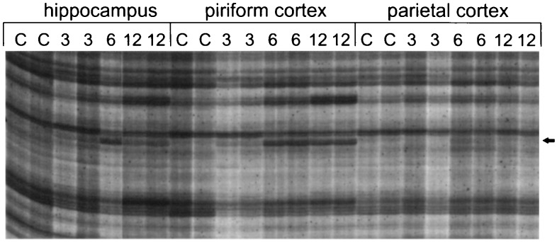
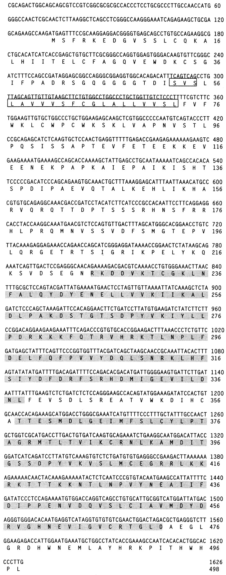
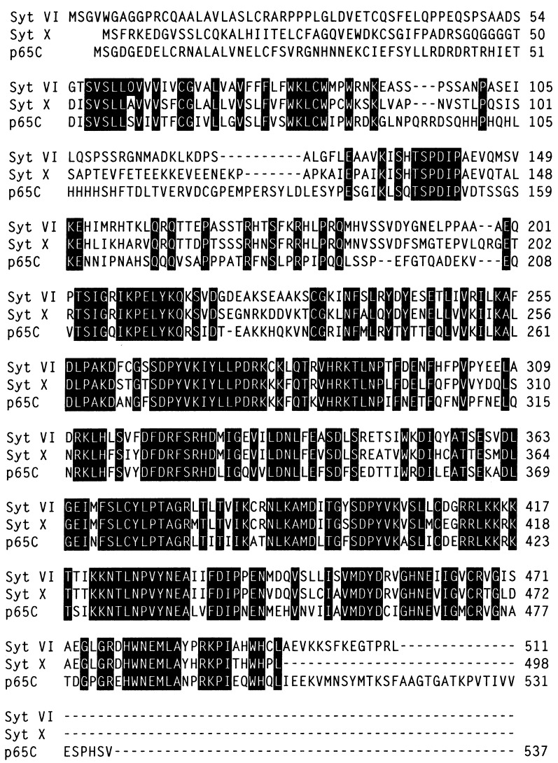
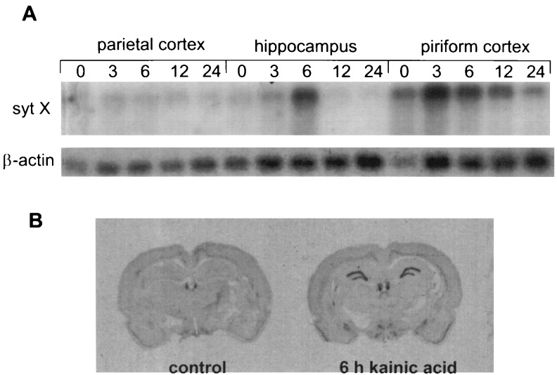
Systemic administration of kainic acid, a cyclic analogue of glutamate, produces many of the clinical features of human temporal lobe epilepsy and status epilepticus in rats, including the induction of motor convulsions and the degeneration of neurons in the hippocampus and piriform cortex. Differential display PCR was used to identify mRNAs that are differentially expressed between degenerating and nondegenerating tissues in the brain after kainic acid-induced seizure activity. A novel cDNA fragment expressed in the degenerating hippocampus and piriform cortex, but not in the nondegenerating parietal cortex, was identified, cloned, and sequenced. This novel cDNA fragment identified a new member of the synaptotagmin gene family that is rapidly and transiently induced in response to seizure activity. Differential expression of this synaptotagmin gene, syt X, was confirmed by Northern blot analysis and in situ hybridization. This novel, inducible synaptotagmin gene may provide a direct link between seizure-induced neuronal gene expression and subsequent modulation of synaptic structure and function.


Figures

Figure 1
Figure 1
Differential display of mRNA comparing gene expression among hippocampus, piriform cortex, and parietal cortex after kainic acid administration. Only a portion of the autoradiogram is shown. C, 3, 6, and 12 denote control, 3 h, 6 h, and 12 h, respectively. Differential display reactions were performed in duplicate using 5′-TTTTTTTTTTTCG-3′ as an anchored primer and 5′-TACAACGAGG-3′ as a random arbitrary primer. The arrow indicates a reproducible PCR-amplified cDNA fragment that appears to be induced only in degenerating tissues (hippocampus and piriform cortex) at 6 and 12 h after kainic acid injection. In the hippocampus and piriform cortices, baseline levels of expression are seen in control animals and at the 3-h time point. By comparison, only baseline levels of expression are observed in the parietal cortex, a tissue in which seizure-induced neuronal degeneration is not observed.
Figure 2
Figure 2
Nucleotide and predicted amino acid sequence of synaptotagmin X. Single letter amino acid codes are used, numbering from the first in-phase ATG codon. The putative transmembrane domain is boxed, and the two C2 domains are indicated by shading. Sequence information from a small portion of the C-terminal tail of syt X is incomplete.
Figure 3
Figure 3
Alignment of rat and marine ray predicted synaptotagmin amino acid sequences. The amino acid sequences of syt VI and syt X from rat and p65C from marine ray are aligned. Identical amino acid residues for all three genes are highlighted in black. The predicted syt X protein is most similar to syt VI and p65C, with overall identities of 62% and 52%, respectively. These three synaptotagmin genes share an additional region of amino acid homology that is not found in other synaptotagmin family members. This region is located between the transmembrane domain and the two C2 domains.
Figure 4
Figure 4
(A) Northern blot analysis of syt X expression. Poly(A)+ RNA from parietal cortex, hippocampus, and piriform cortex at 0, 3, 6, 12, and 24 h after kainic acid-induced seizures was probed with syt X. The probe identifies a single message that is 8.0 kb in length. The blot was also probed with a β-actin oligonucleotide for the purpose of quantitation. (B) In situ hybridization of rat brain sections using an oligonucleotide probe specific for the syt X gene. Coronal section control and 6-h kainic acid-treated rat brain. Examination of control and kainic acid-treated animals revealed that syt X is expressed in the dentate granule cells of the hippocampus and layer II of the piriform cortex.

References

    1. Liang P, Pardee A B. Science. 1992;275:967–971. - PubMed
    1. Liang P, Averboukh L, Pardee A B. Nucleic Acids Res. 1993;21:3269–3275. - PMC - PubMed
    1. Olney J W, Rhee V, Ho O L. Brain Res. 1974;77:507–512. - PubMed
    1. Nadler J V. Life Sci. 1981;29:2031–2042. - PubMed
    1. Sperk G. Prog Neurobiol. 1994;42:1–32. - PubMed

Publication types

MeSH terms

Associated data