. 1997 Mar 18;94(6):2642-7.
doi: 10.1073/pnas.94.6.2642.

Transcription factor NF-kappaB is activated in primary neurons by amyloid beta peptides and in neurons surrounding early plaques from patients with Alzheimer disease

Affiliations

Transcription factor NF-kappaB is activated in primary neurons by amyloid beta peptides and in neurons surrounding early plaques from patients with Alzheimer disease

B Kaltschmidt et al. Proc Natl Acad Sci U S A. .

Abstract

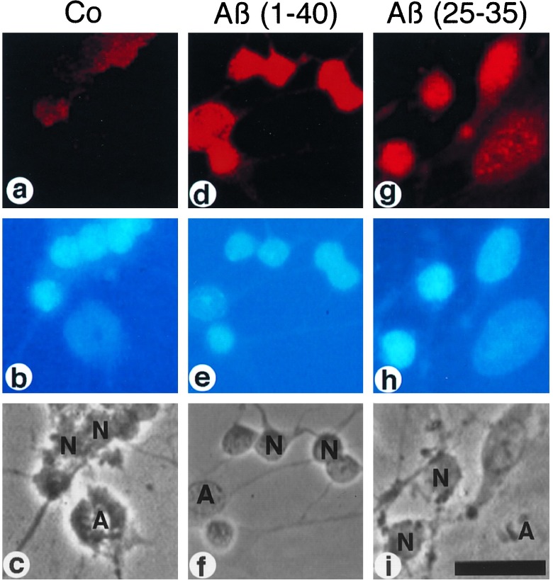
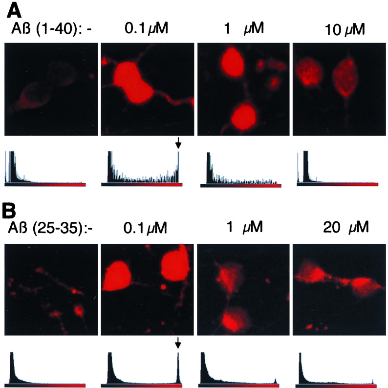
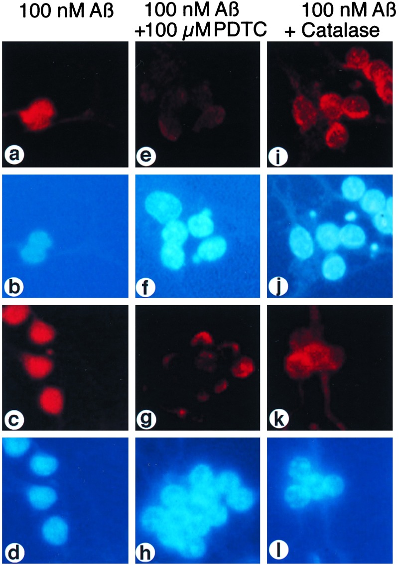
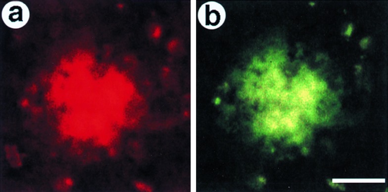
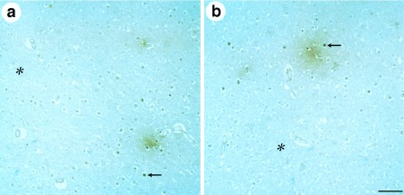
Amyloid beta peptide (A beta)-containing plaques are a hallmark of Alzheimer disease. Here, we show that the neurotoxic A beta, a major plaque component, is a potent activator of the transcription factor NF-kappaB in primary neurons. This activation required reactive oxygen intermediates as messengers because an antioxidant prevented A beta-induced NF-kappaB activation. Maximal activation of NF-kappaB was found with 0.1 microM A beta-(1-40) and 0.1 microM A beta-(25-35) fragments, making a role for NF-kappaB in neuroprotection feasible. Using an activity-specific mAb for the p65 NF-kappaB subunit, activation of NF-kappaB also was observed in neurons and astroglia of brain sections from Alzheimer disease patients. Activated NF-kappaB was restricted to cells in the close vicinity of early plaques. Our data suggest that the aberrant gene expression in diseased nervous tissue is at least in part due to A beta-induced activation of NF-kappaB, a potent immediate-early transcriptional regulator of numerous proinflammatory genes.


Figures

Figure 1
Figure 1
Activation of NF-κB by Aβ in rat cerebellar granule cells. Primary neuronal cells were treated with 100 nM Aβ-(1–40) (d-f) and Aβ-(25–35) (g-i) for 45 min or left untreated (a-c). (a, d, g) Indirect immunofluorescence analysis of cell cultures for p65 NF-κB immunoreactivity using the activity-specific α-p65 mAb (28) and a Cy3-conjugated second antibody (see Materials and Methods). (b, e, h) DNA staining of nuclei in cell cultures using DAPI. (c, f, i) Phase contrast micrographs. N, neurons; A, astrocytes. (Bar = 50 μm.)
Figure 2
Figure 2
Concentration dependence of Aβ-induced NF-κB activation in cerebellar granule cells. (A) Granule cells treated with Aβ-(1–40). (B) Granule cells treated with Aβ-(25–35). (Upper) Indirect immunofluorescence analysis of primary cell cultures for binding of α-p65 mAb. Cells were stimulated for 45 min with the indicated concentrations of Aβ-(1–40). Sections with two to four neurons are shown. Neurons were identified by DAPI staining and by their morphology upon phase contrast microscopy. Analysis of the immunofluorescence intensity of a larger area of the specimen in one optical plane. Histograms for each concentration are shown. The ordinate (linear scale) depicts relative amounts of pixels. The fluorescence intensities of the Cy3 chromophor are shown on a linear scale on the abscissa. Arrows indicate the maximal signals obtained at 100 nM Aβ peptides.
Figure 3
Figure 3
The effect of the antioxidant PDTC and catalase on Aβ-induced NF-κB activation. Granule cells were incubated for 45 min with 100 nM Aβ alone [a and b, Aβ-(1–40); c and d, Aβ-(25–35)] or with Aβ and 100 μM PDTC, which was added to cell cultures 10 min before Aβ [e and f, Aβ-(1–40); g and h, Aβ-(25–35)], or with 100 nM Aβ plus 400 μg/ml catalase, which was added to cells 1 h before the Aβ peptide [i and j, Aβ-(1–40); k and l, Aβ-(25–35)]. Cell cultures were analyzed by indirect immunofluorescence for an increase of α-p65 mAb immunoreactivity (a, e, i, c, g, k) and by nuclear DAPI-staining (b, f, j, d, h, l).
Figure 4
Figure 4
Colocalization of α-p65 mAb and anti-Aβ immunoreactivities in senile plaques from brains of AD patients. In double immunofluorescence experiments, cryosections from patients with AD were immunostained with polyclonal anti-Aβ (b) and monoclonal anti-p65 (a) antibodies. Bound antibodies were detected by species-specific second antibodies conjugated to either Cy3 [red fluorescence, anti-p65 (a)] or dichlorotriazinyl amino fluorescein [green fluorescence, anti-Aβ (b)]. (Bar = 10 μm.)
Figure 5
Figure 5
Immunohistochemical analysis of plaques in cortical sections of AD brains for α-p65 mAb immunoreactivity. Plaques are visualized by immunoreactivity (brown color). Examples of α-p65 immunoreactive cell nuclei (arrows) and surrounding plaques are depicted. Different examples for primitive plaque types are shown. Regions distant from plaques have reduced α-p65 immunoreactivity (asterisks). (Bar = 50 μm.)

References

    1. Glenner G G, Wong C W. Biochem Biophys Res Commun. 1984;120:885–890. - PubMed
    1. Masters C L, Simms G, Weinmann N A, Multhaup G, McDonald B L, Beyreuther K. Proc Natl Acad Sci USA. 1985;82:4245–4249. - PMC - PubMed
    1. Kang J, Lemaire H G, Unterbeck A, Salbaum M, Masters C L, Grzeschik K H, Multhaup G, Beyreuther K, Müller-Hill B. Nature (London) 1987;325:733–736. - PubMed
    1. Müller U, Cristina N, Li Z W, Wolfer D P, Lipp H P, Rulicke T, Brandner S, Aguzzi A, Weissmann C. Cell. 1994;79:755–765. - PubMed
    1. Zheng H, Jiang M, Trumbauer M E, Sirinahsinghji D J S, Hopkins R, et al. Cell. 1995;81:525–531. - PubMed

Publication types

MeSH terms