. 1997 Mar 18;94(6):2648-53.
doi: 10.1073/pnas.94.6.2648.

Neuritin: a gene induced by neural activity and neurotrophins that promotes neuritogenesis

Affiliations

Neuritin: a gene induced by neural activity and neurotrophins that promotes neuritogenesis

G S Naeve et al. Proc Natl Acad Sci U S A. .

Abstract

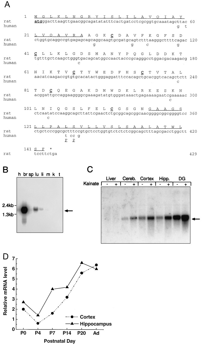
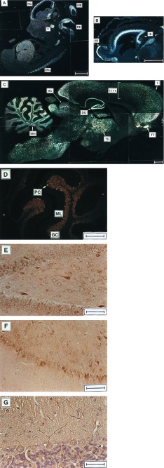
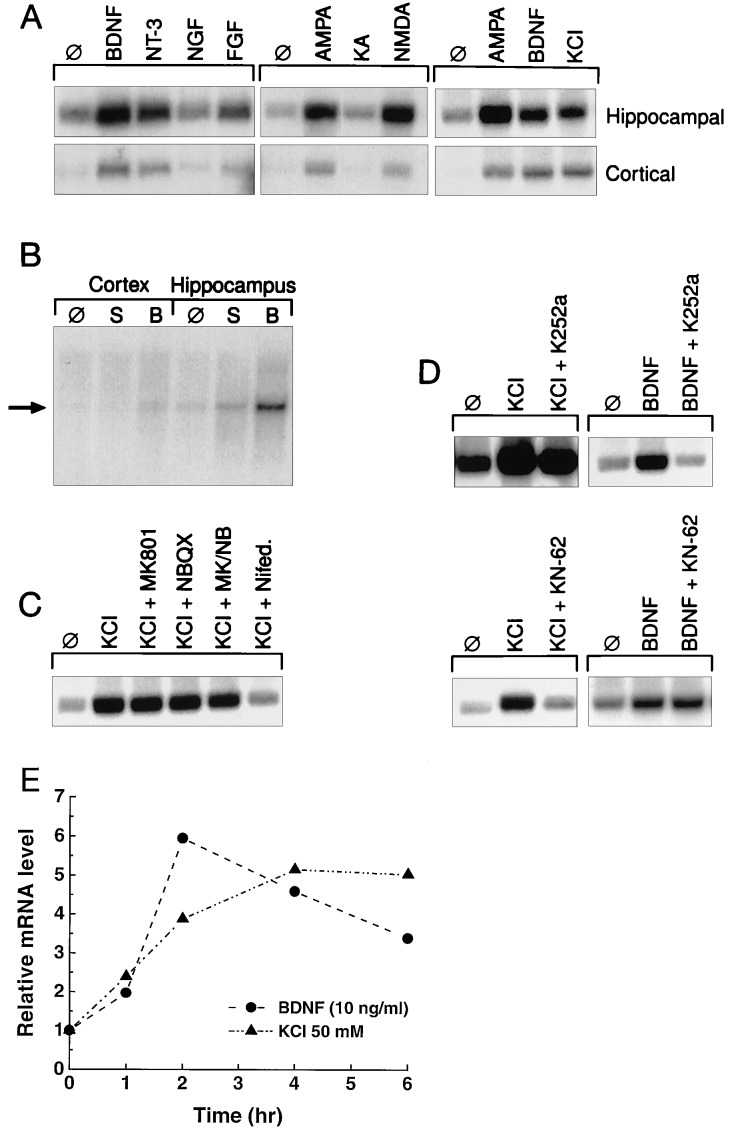
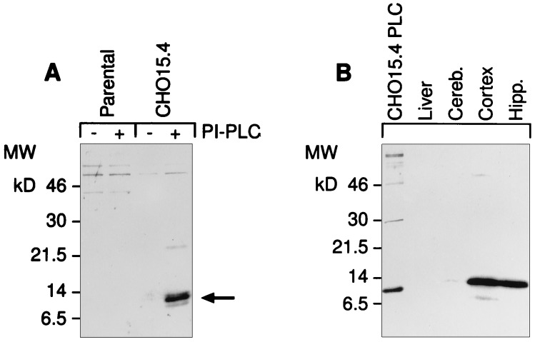
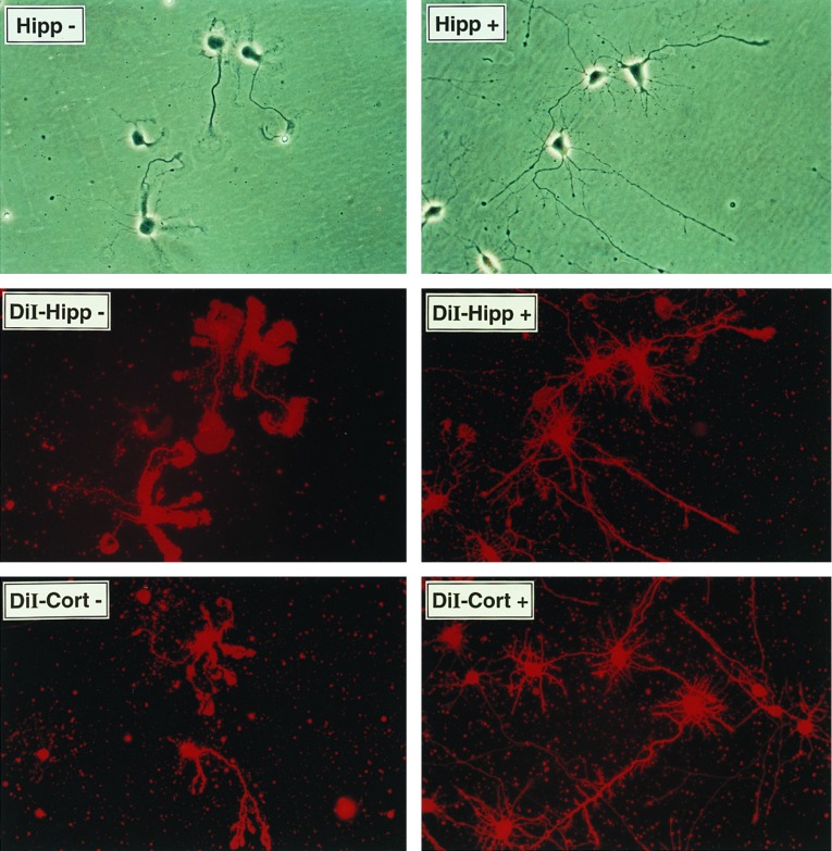
Neural activity and neurotrophins induce synaptic remodeling in part by altering gene expression. A cDNA encoding a glycosylphoshatidylinositol-anchored protein was identified by screening for hippocampal genes that are induced by neural activity. This molecule, named neuritin, is expressed in postmitotic-differentiating neurons of the developing nervous system and neuronal structures associated with plasticity in the adult. Neuritin message is induced by neuronal activity and by the activity-regulated neurotrophins BDNF and NT-3. Purified recombinant neuritin promotes neurite outgrowth and arborization in primary embryonic hippocampal and cortical cultures. These data implicate neuritin as a downstream effector of activity-induced neurite outgrowth.


Figures

Figure 1
Figure 1
Deduced amino sequence and expression profile of neuritin cDNA. (A) The full sequence of rat neuritin is shown. The coding region of human neuritin is identical to the rat sequence in all but 14 bp. Only the nucleotides and amino acids that differ from the rat sequence are shown. The predicted signal peptides for secretion and GPI anchoring are underlined. The six cysteine residues of the mature protein are in boldface type. (B) Multiple-tissue Northern blot of 10 μg of poly(A)+ RNA from human tissues probed with a 32P-labeled cRNA probe of neuritin coding sequence. The neuritin message migrates between 1.6 and 2.0 kb. h, Heart; br, brain; sp, spleen; lu, lung; li, liver; m, muscle; k, kidney; t, testis. (C) Neuritin mRNA is present in different regions of the brain, and the expression is stimulated by KA in vivo. Northern blot of total RNA (10 μg) isolated from control or KA-treated rats (i.p. injection, 10 mg/kg for 6 hr) probed with the neuritin cRNA probe. Cereb., cerebellum; Hipp., hippocampus; DG, dentate gyrus. (D) Quantitated Northern blot analysis results of RNA harvested at different postnatal time points. Ad, adult (8 weeks).
Figure 2
Figure 2
Expression of embryonic neuritin mRNA and protein in situ. Neuritin mRNA is detected with a 32P-labeled neuritin cRNA probe in parasaggital sections of rat embryos. (A) Whole embryo, E15. NC, neocortex; ME, mesencephalon; RE, rhombencephalon; V, trigeminal ganglion; DRG, dorsal root ganglia. (B) E18 brain. Mi, mitral cell layer of olfactory bulb; Hi, hippocampus. (C and D) Adult rat brain. High levels of expression are observed in cortical layers II–VI, pyramidal and granule cells (DG) of the hippocampal formation, thalamus (TH), superior colliculus (SC), tenia tecta (TT), middle medial nucleus (MM), and the granule cell layer (GC) and scattered Purkinje cells of the cerebellum. ML, molecular layer. Anti-neuritin antisera on parasaggital sections of adult rat brain labels cells in DG (E) and subiculum/CA1 (F) of the hippocampus and Purkinje cells (G) of the cerebellum. Neuritin expression is detected with polyclonal antisera generated against whole recombinant (E and F) or a peptide fragment of neuritin (G). Immunopositive cells have an orange-brown appearance. [Bars = 1 mm (AC), 500 μm (D), 100 μm (E and F), and 50 μm (G).]
Figure 3
Figure 3
Regulation of neuritin mRNA. (A) Neuritin gene expression is induced in primary hippocampal or cortical neurons by NTs, glutamate analogs, and depolarization by KCl (10 ng/ml NTs/growth factors, 30 μM glutamate analogs, and 50 mM KCl). Total RNA was harvested from primary embryonic rat neurons (E18, 7 days in vitro, treated for 6 hr) and used in Northern blot analysis (5 μg). (B) Intracranial injection of BDNF into postnatal day 4 rats induces neuritin mRNA in vivo. Total RNA from the indicated brain tissues was isolated after intraventricular injection. Ten micrograms of total RNA was analyzed by Northern blot analysis. ∅, No treatment; S, saline injection; B, BDNF. (C) Induction of neuritin mRNA by KCl occurs through activation of voltage-gated calcium channels in hippocampal neurons. (D and E) BDNF and KCl induce neuritin message in hippocampal neurons independently and with distinct kinetics. The inhibitors (NMDA, 10 μM MK-801; AMPA, 10 μM NBQX; voltage-gated calcium channels, 5 μM nifedipine; TrkB, 50 nM K252a; α-CAMKII, 10 μM KN-62) were added to cultures 30 min before induction.
Figure 4
Figure 4
Neuritin is a GPI-anchored protein that promotes neuritogenesis in primary hippocampal and cortical cells. (A) Recombinant human neuritin is sensitive to PI-PLC. Stably transfected CHO cells carrying the neuritin expression vector or the empty parental vector were treated with PI-PLC release buffer alone (−) or in the presence (+) of PI-PLC (0.4 unit/ml). Supernatants were analyzed by probing Western blots with affinity-purified antisera to a synthetic peptide derived from the C-terminal part of mature neuritin. (B) Endogenous neuritin is solubilized in Triton X-114. Tissues were harvested from control or KA-treated rats (i.p., 10 mg/kg, 8 hr) and proteins were extracted by phase separation with Triton X-114. Proteins were analyzed as in A. The first lane is PI-PLC extract from CHO cells expressing recombinant human neuritin (CHO15 cells). The difference in migration is due to the altered mobility of proteins containing an intact GPI anchor (Triton extracts). (C) Histidine-tagged neuritin produced in CHO cells and purified from conditioned media by nickel chromatography promotes neuritogenesis in embryonic hippocampal and cortical cells. Primary neurons from E18 rats were plated at 5 × 10³ cells per square centimeter on poly-l-lysine-coated culture dishes in the presence of 150 ng/ml neuritin (+), or a corresponding control nickel-column fraction of parental vector CHO-conditioned media (−). The factor was added at the time of plating and neurite outgrowth was monitored daily. On day 4 of culture, cells were nonspecifically labeled with the lipophilic dye DiI (10 μM in dimethyl sulfoxide) and photographed using phase contrast (Top) or fluorescent (Middle and Bottom) microscopy.
Figure 4
Figure 4
Neuritin is a GPI-anchored protein that promotes neuritogenesis in primary hippocampal and cortical cells. (A) Recombinant human neuritin is sensitive to PI-PLC. Stably transfected CHO cells carrying the neuritin expression vector or the empty parental vector were treated with PI-PLC release buffer alone (−) or in the presence (+) of PI-PLC (0.4 unit/ml). Supernatants were analyzed by probing Western blots with affinity-purified antisera to a synthetic peptide derived from the C-terminal part of mature neuritin. (B) Endogenous neuritin is solubilized in Triton X-114. Tissues were harvested from control or KA-treated rats (i.p., 10 mg/kg, 8 hr) and proteins were extracted by phase separation with Triton X-114. Proteins were analyzed as in A. The first lane is PI-PLC extract from CHO cells expressing recombinant human neuritin (CHO15 cells). The difference in migration is due to the altered mobility of proteins containing an intact GPI anchor (Triton extracts). (C) Histidine-tagged neuritin produced in CHO cells and purified from conditioned media by nickel chromatography promotes neuritogenesis in embryonic hippocampal and cortical cells. Primary neurons from E18 rats were plated at 5 × 10³ cells per square centimeter on poly-l-lysine-coated culture dishes in the presence of 150 ng/ml neuritin (+), or a corresponding control nickel-column fraction of parental vector CHO-conditioned media (−). The factor was added at the time of plating and neurite outgrowth was monitored daily. On day 4 of culture, cells were nonspecifically labeled with the lipophilic dye DiI (10 μM in dimethyl sulfoxide) and photographed using phase contrast (Top) or fluorescent (Middle and Bottom) microscopy.

References

    1. Bailey C H, Kandel E R. Annu Rev Physiol. 1993;55:397–426. - PubMed
    1. Bonhoeffer T. Curr Opin Neurobiol. 1996;6:119–126. - PubMed
    1. McNaughton B L. Annu Rev Physiol. 1993;55:375–396. - PubMed
    1. Thoenen H. Science. 1995;270:593–598. - PubMed
    1. Qian Z, Gilbert M E, Colicos M A, Kandel E R, Kuhl D. Nature (London) 1993;361:453–457. - PubMed

MeSH terms