. 1997 Mar 18;94(6):2671-5.
doi: 10.1073/pnas.94.6.2671.

Dual activity maps in primate visual cortex produced by different temporal patterns of zif268 mRNA and protein expression

Affiliations

Dual activity maps in primate visual cortex produced by different temporal patterns of zif268 mRNA and protein expression

A Chaudhuri et al. Proc Natl Acad Sci U S A. .

Abstract

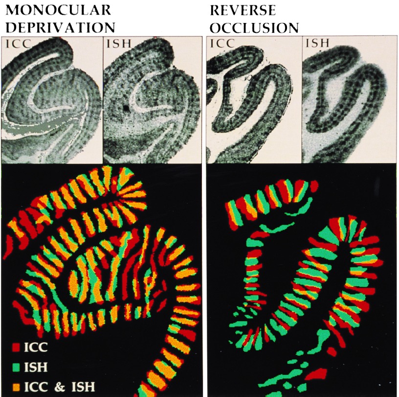
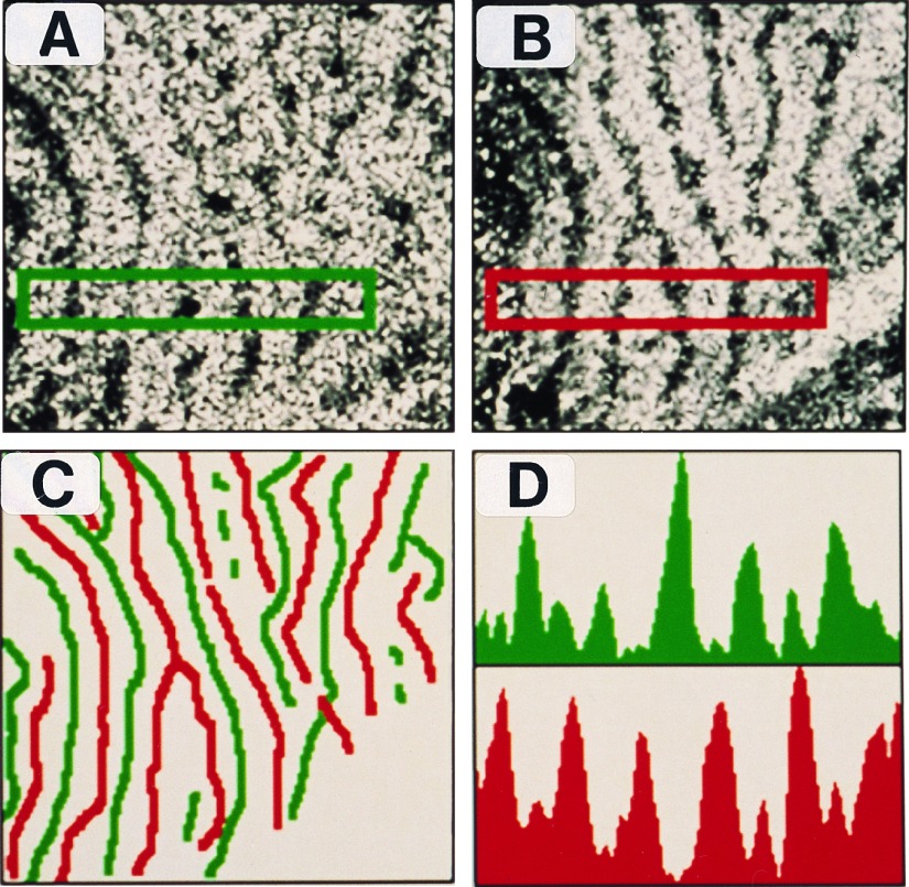
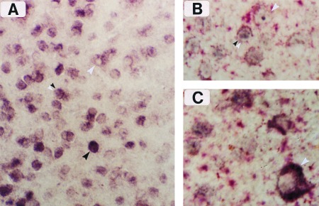
The inducible nature of the immediate-early genes (IEGs) c-fos and zif268 allows their products to be used as activity markers in the brain. The utility of such markers in general is restricted because they can resolve only neurons activated by a single stimulus. To overcome this limitation, we have developed a double-label technique that exploits the dissimilar time course of zif268 mRNA and protein induction, allowing them to be separately induced by two different stimuli and independently stained to provide a visual display of neurons that are responsive to each stimulus. Two powerful features of this new imaging technique-the possibility of staining separate populations of activated neurons and the ability to visualize them at the cellular level-should extend IEG applications in biological activity mapping.


Figures

Figure 1
Figure 1
Visualization of OD columns in coronal sections of area V1 by Zif268 mRNA and protein staining. (Upper) ICC and ISH staining results from adjacent sections. (×2.2.) (Lower) The OD columns were delineated, color-coded, and digitally superimposed. (×3.8.) The protein-positive and mRNA-positive regions show significant overlap in the MD condition, while they are largely complementary in the RO condition.
Figure 2
Figure 2
Topographic organization of mRNA- and protein-stained ocular dominance columns in flattened views of area V1. (A) Film autoradiogram showing OD columns labeled by zif268 mRNA. (×7.2.) (B) Immunostained section showing OD columns labeled by Zif268 protein. (×7.2.) (C) Topographic layout of zif268 mRNA (green) and protein (red) labeled columns shows that they have a complementary, nonoverlapping arrangement. (×7.2.) (D) Optical density profiles confined to the rectangular region in the stained sections show that regions of intense mRNA labeling are accompanied by poor protein staining and vice versa.
Figure 3
Figure 3
Cellular resolution of zif268 mRNA and protein labeling by simultaneous ISH (dig-conjugated complementary RNA probe) and immunostaining. (A) Border region of two OD columns in layer II, showing single-labeled cells that are mRNA-positive (white arrowhead) or protein-positive (black arrowhead). Double-labeled cells are also visible in this figure (double arrowhead), and they are enhanced when differential color staining is applied, as in B and C. (×250.) (B) Immunoreactive product (blue) and dig-mRNA complex (red) can be seen together in several double-labeled cells from layer II. (×400.) (C) Meynert cell from layer VI (white arrowhead) enriched with dig-conjugated reaction product surrounding a negatively stained nucleus. (×400.)

References

    1. Cole A J, Saffen D W, Baraban J M, Worley P F. Nature (London) 1989;340:474–476. - PubMed
    1. Sheng M, Greenberg M E. Neuron. 1990;4:477–485. - PubMed
    1. Morgan J I, Curran T. Annu Rev Neurosci. 1991;14:421–451. - PubMed
    1. Ghosh A, Greenberg M E. Science. 1995;268:239–247. - PubMed
    1. Lerea L S, Carlson N G, McNamara J O. Mol Pharmacol. 1995;47:1119–1125. - PubMed

Publication types

LinkOut - more resources