. 1997 Apr 21;137(2):275-89.
doi: 10.1083/jcb.137.2.275.

Structural protein 4.1 in the nucleus of human cells: dynamic rearrangements during cell division

Affiliations

Structural protein 4.1 in the nucleus of human cells: dynamic rearrangements during cell division

S W Krauss et al. J Cell Biol. .

Abstract

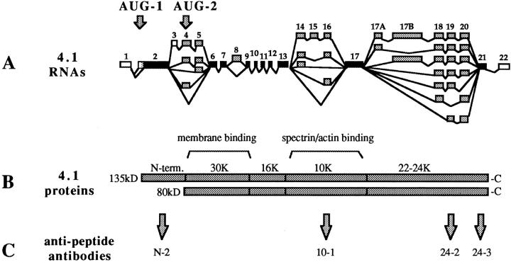
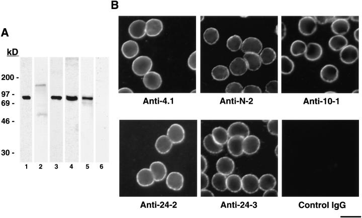
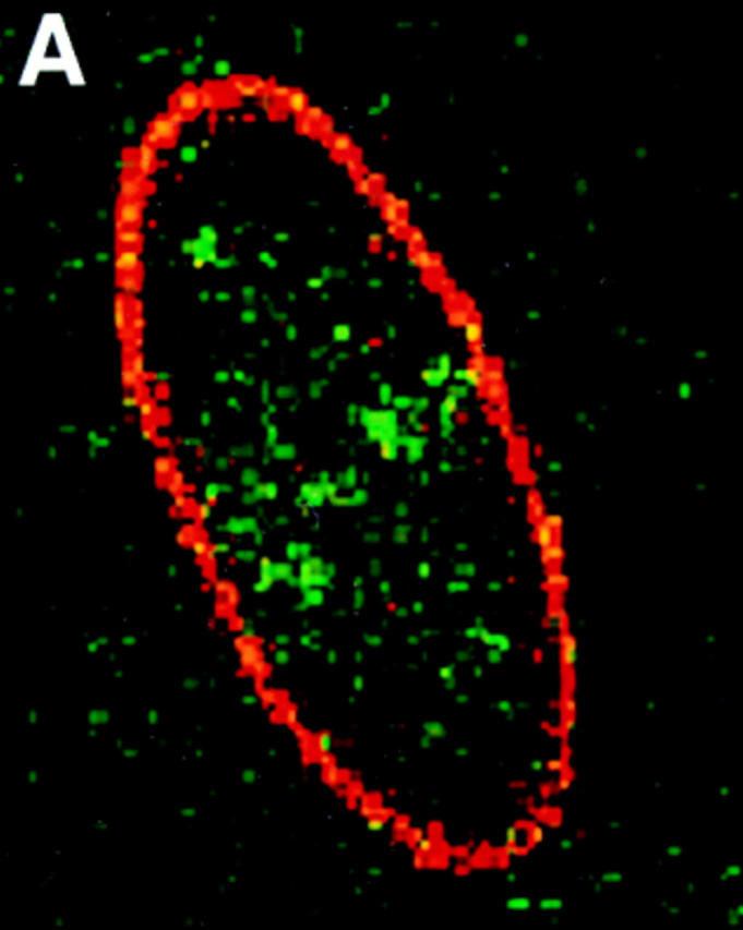
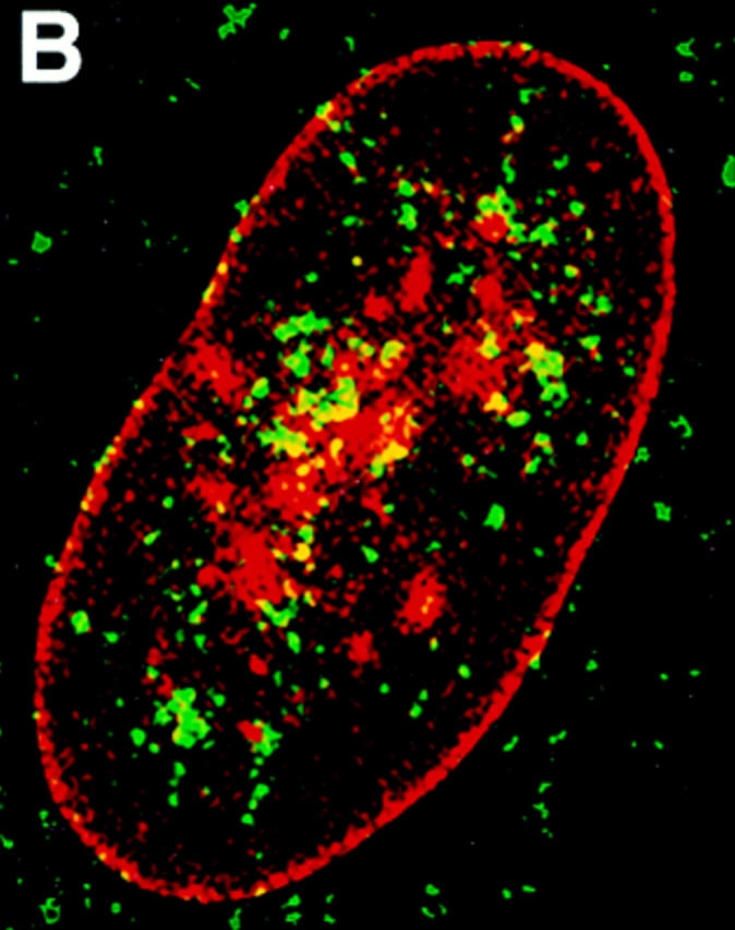
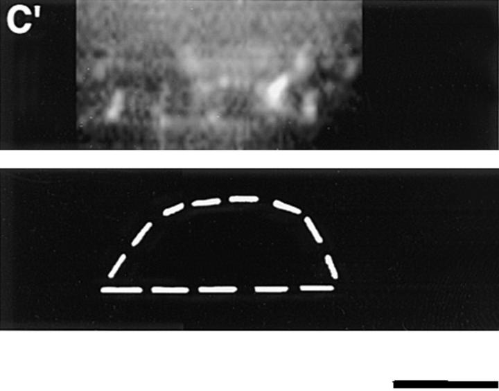
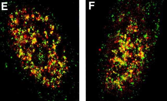
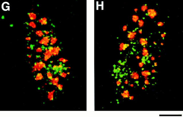
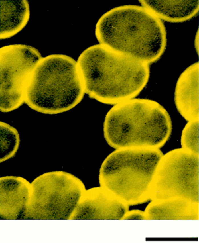
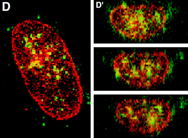
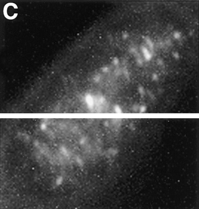
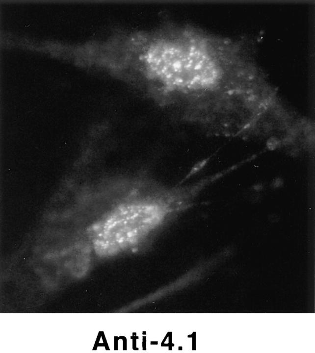
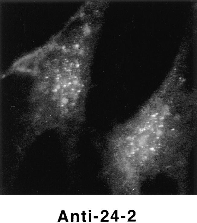
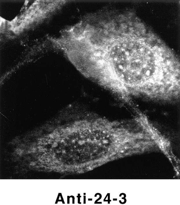
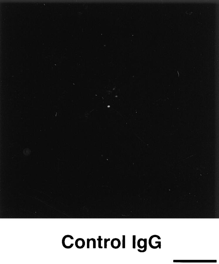
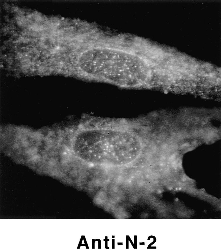
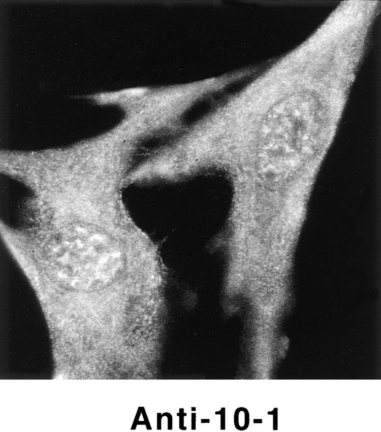
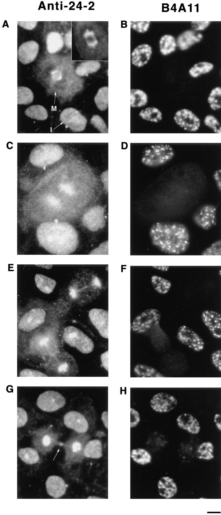
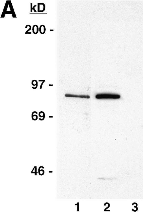
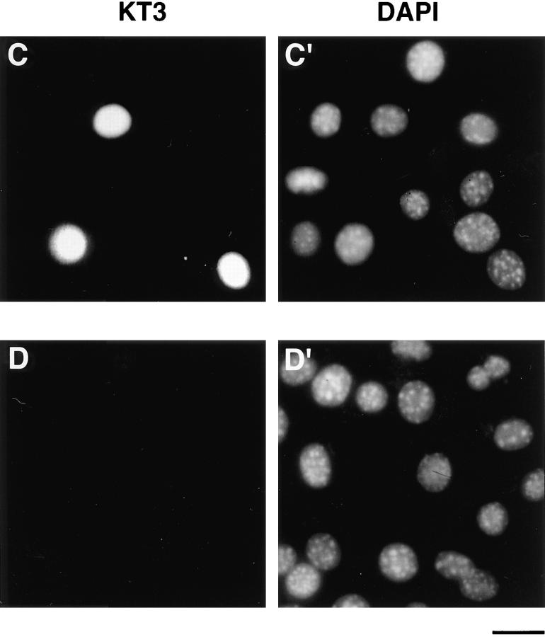
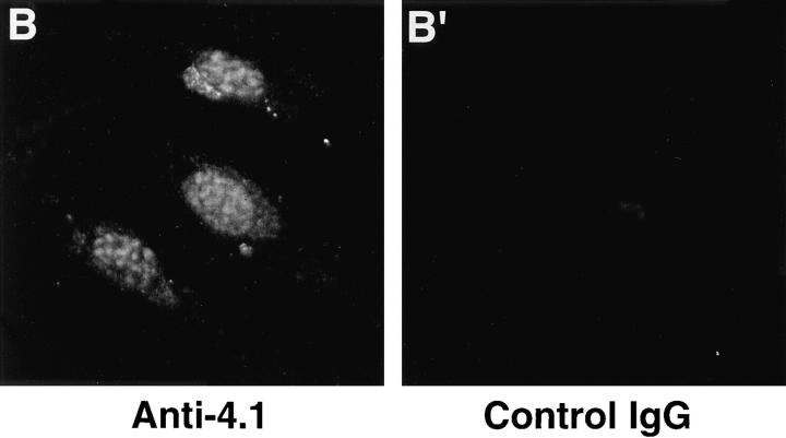
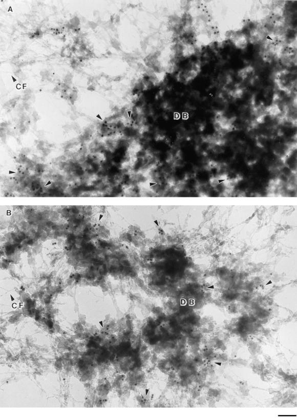
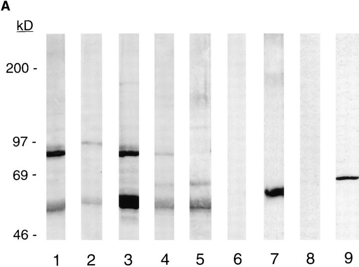
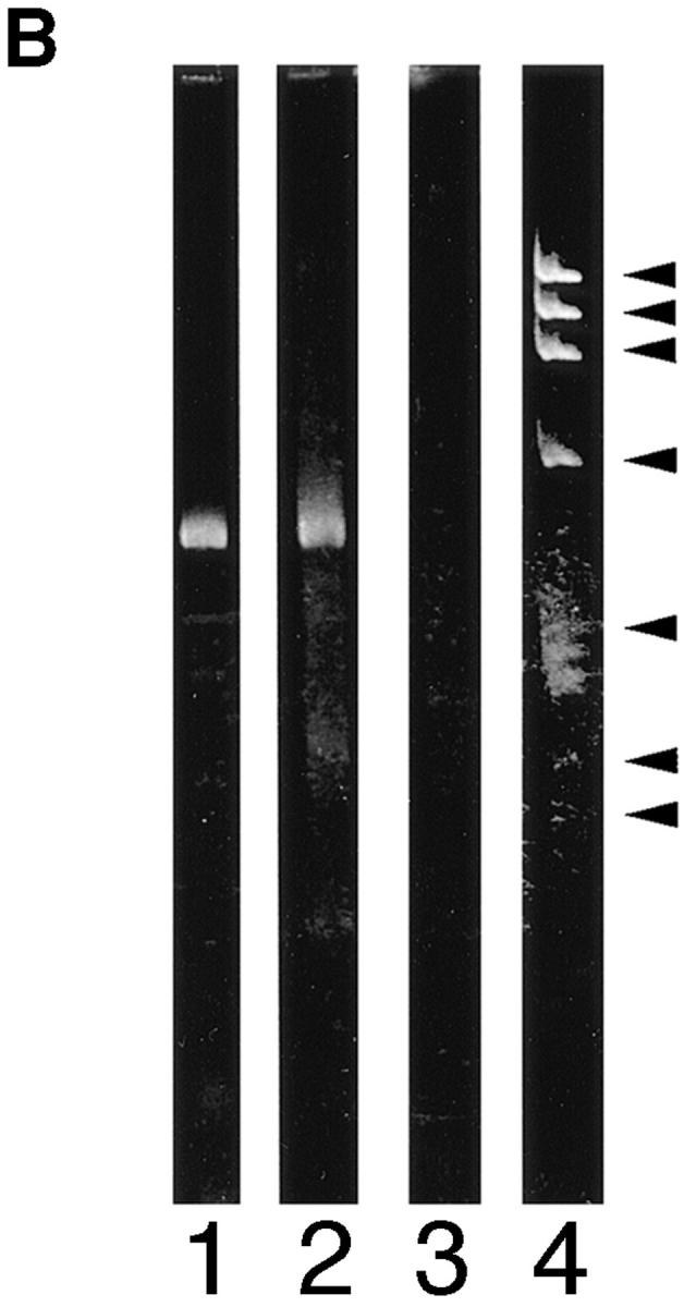
Structural protein 4.1, first identified as a crucial 80-kD protein in the mature red cell membrane skeleton, is now known to be a diverse family of protein isoforms generated by complex alternative mRNA splicing, variable usage of translation initiation sites, and posttranslational modification. Protein 4.1 epitopes are detected at multiple intracellular sites in nucleated mammalian cells. We report here investigations of protein 4.1 in the nucleus. Reconstructions of optical sections of human diploid fibroblast nuclei using antibodies specific for 80-kD red cell 4.1 and for 4.1 peptides showed 4.1 immunofluorescent signals were intranuclear and distributed throughout the volume of the nucleus. After sequential extractions of cells in situ, 4.1 epitopes were detected in nuclear matrix both by immunofluorescence light microscopy and resinless section immunoelectron microscopy. Western blot analysis of fibroblast nuclear matrix protein fractions, isolated under identical extraction conditions as those for microscopy, revealed several polypeptide bands reactive to multiple 4.1 antibodies against different domains. Epitope-tagged protein 4.1 was detected in fibroblast nuclei after transient transfections using a construct encoding red cell 80-kD 4.1 fused to an epitope tag. Endogenous protein 4.1 epitopes were detected throughout the cell cycle but underwent dynamic spatial rearrangements during cell division. Protein 4.1 was observed in nucleoplasm and centrosomes at interphase, in the mitotic spindle during mitosis, in perichromatin during telophase, as well as in the midbody during cytokinesis. These results suggest that multiple protein 4.1 isoforms may contribute significantly to nuclear architecture and ultimately to nuclear function.


Figures

Figure 1
Figure 1
Protein 4.1 peptide domains used to generate antibodies. (A) A schematic diagram of protein 4.1 mRNAs displaying multiple combinations of splicing pathways possible among 4.1 alternative exons. In this format, exons are coded as follows: solid bars, constitutive; shaded bars, alternative; open bars, noncoding. The arrows on top indicate the positions of alternative translation initiation sites, AUG-1 and AUG-2. The figure is derived from Conboy et al. (1991). (B) Examples of protein 4.1 isoforms derived from different translation initiation sites. The 80-kD prototypical red cell isoform is produced from AUG-2 and can be present in nucleated and nonnucleated cells. Chymotryptic fragments of this isoform include a 30-kD membrane binding domain, a 16-kD domain, a 10-kD spectrin and actin binding domain, and the 22–24-kD domain (Leto and Marchesi, 1984). Higher molecular mass 4.1 isoforms, present in nucleated cells, use AUG-1 to generate an additional 209–amino acid “NH2-terminal extension” (N-term). (C) Synthetic peptides derived from the 4.1 amino acid sequence at the positions indicated by the arrows were used to immunize rabbits. IgGs were prepared from N-2, 10-1, 24-2, and 24-3 sera by affinity purification using the homologous peptide. The sequences for the peptides are given in the Methods section.
Figure 2
Figure 2
Characterization of antibodies directed against red cell 80-kD 4.1 and 4.1 peptide domains. (A) Human red cell membrane fractions (Conboy et al., 1993) were separated by SDS-PAGE, transferred to nitrocellulose, and incubated with the following IgGs: lane 1, anti-RBC 4.1; lane 2, anti–N-2; lane 3, anti–10-1; lane 4, anti–24-2; lane 5, anti–24-3; lane 6, control IgG. Protein 4.1 in red cells migrates at ∼80 kD. Although this is the predominant 4.1 isoform in red cells, a faint band at ∼135 kD detected by anti–N-2 (lane 2) is also detected by other 4.1 IgGs when increased amounts of red cell membrane preparations are applied to gels (Conboy et al., 1991). (B) Human red cells were fixed by methanol and probed by indirect immunofluorescence using primary antibodies as indicated and FITCconjugated secondary antibodies. The faint staining by anti–N-2 most likely reflects the small amount of ∼135-kD 4.1 also detected by Western blot analysis of red cell membranes. Bar, 10 μm.
Figure 5
Figure 5
Distribution of protein 4.1 epitopes in the interphase nucleus of human fibroblasts visualized by immunofluorescence. Location of the nucleus was confirmed in all cases by DAPI staining (not shown). (A and B) Double-label confocal microscopy of cells stained by protein 4.1 antibody 24-2 (green) and (A) antibody against nuclear pores (red) or (B) lamin B (red). The majority of nuclear protein 4.1 foci are interior to the periphery of the nucleus as demarcated by immunofluorescence of pores within nuclear membrane and by its subadjacent network of lamin B fibers. (Additional lamin B sites in the nuclear interior have been reported [Bridger et al., 1993; Moir et al., 1994]). Similar results were obtained by imaging epitopes for protein 4.1 antibodies 24-3, 10-1, and N-2 relative to antipore and antilamin epitopes. (C) Three-dimensional image analysis of nuclear epitopes probed with anti-RBC 80-kD 4.1 showing a 0.3-μm horizontal optical midsection of an immunofluorescent cell with a line indicating the position of the vertical reconstruction depicted in C′. Since optical resolution in the z-axis (vertical) is less than in the x-y (horizontal) axis, the reconstructed image is less sharp; the dome shape of the top of the cell and the flat bottom plane of the coverslip are somewhat obscured by the coincidence of out of focus light. (A cartoon depicting the approximate shape of the vertical cross-section through the nucleus is presented in the bottom panel.) Thus, it is apparent that epitopes for protein 4.1 lie in numerous planes within the volume of the cell. Similar results were obtained in three-dimensional reconstructions using the 24-2, 24-3, and 10-1 antibodies as probes. (D and D′) Double-label confocal microscopy of a fibroblast using anti–24-2 (green) and anti-NuMA (red). In a horizontal plane through the midsection of the cell (D), protein 4.1 foci appeared within the NuMA-stained nuclear interior. (D′) Three-dimensional reconstructions were made of three different vertical planes from this NuMA:24-2 double-labeled cell. The flattened plane of the coverslip appears at the top of the vertical reconstructed images. The conclusion that 4.1 epitopes localized at multiple planes throughout the volume of the nucleus is entirely consistent with the observations obtained in images A, B, and C. The same conclusion was reached when cells were probed with antibodies against NuMA and RBC 80-kD protein 4.1. (E and F) Double-label confocal microscopy of cells probed with antibody 24-2 (green) and antibody against PCNA (red). Optical sections through the fibroblast nucleus show that 24-2 (green) and PCNA (red) epitopes coincided (yellow) in many areas except in the vicinities of the nucleoli, which either are relatively devoid of any signal or sometimes contained a clustering of 24-2 (green) signals. Both PCNA and 24-2 epitopes resided in multiple planes throughout the nucleus (not shown). (G and H) Double-label confocal microscopy of cells probed with antibody 24-2 (green) and antibody against SC-35 (red). Optical sections are in the plane of the cell containing SC-35 domains; most SC-35 domains in fibroblasts are located in a single plane (Carter et al., 1993). It is apparent that the vast majority of the SC-35 domains contained areas of coincident staining with 24-2 (seen as yellow coloration), most often at the periphery of the SC-35 domains. In optical sections above and below SC-35, additional 24-2 signals are observed (not shown), consistent with the conclusions from the localization data presented in A–F. Bars, 3.5 μm (8 μm in the vertical dimension [C′ and D′]).
Figure 5
Figure 5
Distribution of protein 4.1 epitopes in the interphase nucleus of human fibroblasts visualized by immunofluorescence. Location of the nucleus was confirmed in all cases by DAPI staining (not shown). (A and B) Double-label confocal microscopy of cells stained by protein 4.1 antibody 24-2 (green) and (A) antibody against nuclear pores (red) or (B) lamin B (red). The majority of nuclear protein 4.1 foci are interior to the periphery of the nucleus as demarcated by immunofluorescence of pores within nuclear membrane and by its subadjacent network of lamin B fibers. (Additional lamin B sites in the nuclear interior have been reported [Bridger et al., 1993; Moir et al., 1994]). Similar results were obtained by imaging epitopes for protein 4.1 antibodies 24-3, 10-1, and N-2 relative to antipore and antilamin epitopes. (C) Three-dimensional image analysis of nuclear epitopes probed with anti-RBC 80-kD 4.1 showing a 0.3-μm horizontal optical midsection of an immunofluorescent cell with a line indicating the position of the vertical reconstruction depicted in C′. Since optical resolution in the z-axis (vertical) is less than in the x-y (horizontal) axis, the reconstructed image is less sharp; the dome shape of the top of the cell and the flat bottom plane of the coverslip are somewhat obscured by the coincidence of out of focus light. (A cartoon depicting the approximate shape of the vertical cross-section through the nucleus is presented in the bottom panel.) Thus, it is apparent that epitopes for protein 4.1 lie in numerous planes within the volume of the cell. Similar results were obtained in three-dimensional reconstructions using the 24-2, 24-3, and 10-1 antibodies as probes. (D and D′) Double-label confocal microscopy of a fibroblast using anti–24-2 (green) and anti-NuMA (red). In a horizontal plane through the midsection of the cell (D), protein 4.1 foci appeared within the NuMA-stained nuclear interior. (D′) Three-dimensional reconstructions were made of three different vertical planes from this NuMA:24-2 double-labeled cell. The flattened plane of the coverslip appears at the top of the vertical reconstructed images. The conclusion that 4.1 epitopes localized at multiple planes throughout the volume of the nucleus is entirely consistent with the observations obtained in images A, B, and C. The same conclusion was reached when cells were probed with antibodies against NuMA and RBC 80-kD protein 4.1. (E and F) Double-label confocal microscopy of cells probed with antibody 24-2 (green) and antibody against PCNA (red). Optical sections through the fibroblast nucleus show that 24-2 (green) and PCNA (red) epitopes coincided (yellow) in many areas except in the vicinities of the nucleoli, which either are relatively devoid of any signal or sometimes contained a clustering of 24-2 (green) signals. Both PCNA and 24-2 epitopes resided in multiple planes throughout the nucleus (not shown). (G and H) Double-label confocal microscopy of cells probed with antibody 24-2 (green) and antibody against SC-35 (red). Optical sections are in the plane of the cell containing SC-35 domains; most SC-35 domains in fibroblasts are located in a single plane (Carter et al., 1993). It is apparent that the vast majority of the SC-35 domains contained areas of coincident staining with 24-2 (seen as yellow coloration), most often at the periphery of the SC-35 domains. In optical sections above and below SC-35, additional 24-2 signals are observed (not shown), consistent with the conclusions from the localization data presented in A–F. Bars, 3.5 μm (8 μm in the vertical dimension [C′ and D′]).
Figure 3
Figure 3
Detection of nuclear epitopes of protein 4.1 by immunofluorescent light microscopy in WI38 cells. Cultured WI38 human fibroblasts were fixed in acetone for probing with anti-RBC 80-kD 4.1 and N-2, in methanol for probing with anti–24-2, or in formaldehyde for probing with anti–10-1 and anti–24-3, followed by incubation with a FITC-labeled secondary antibody. Punctate nuclear signals, detected with all the protein 4.1 antibodies, were particularly prominent with anti-RBC 80-kD 4.1 and anti–24-2. Localization of FITC signals within the nuclei of all cells was confirmed by comparison to DAPI fluorescence of the cells with 4.1 fluorescence (not shown). Antibodies N-2, 10-1, and 24-3 also produced considerable cytoplasmic staining. Staining patterns were in large part independent of the fixation method. Controls showed no fluorescent patterns when imaged under the same conditions as experimental samples. Bar, 10 μm.
Figure 3
Figure 3
Detection of nuclear epitopes of protein 4.1 by immunofluorescent light microscopy in WI38 cells. Cultured WI38 human fibroblasts were fixed in acetone for probing with anti-RBC 80-kD 4.1 and N-2, in methanol for probing with anti–24-2, or in formaldehyde for probing with anti–10-1 and anti–24-3, followed by incubation with a FITC-labeled secondary antibody. Punctate nuclear signals, detected with all the protein 4.1 antibodies, were particularly prominent with anti-RBC 80-kD 4.1 and anti–24-2. Localization of FITC signals within the nuclei of all cells was confirmed by comparison to DAPI fluorescence of the cells with 4.1 fluorescence (not shown). Antibodies N-2, 10-1, and 24-3 also produced considerable cytoplasmic staining. Staining patterns were in large part independent of the fixation method. Controls showed no fluorescent patterns when imaged under the same conditions as experimental samples. Bar, 10 μm.
Figure 9
Figure 9
Dynamic redistribution of protein 4.1 antigens during the cell cycle. CaSki cells were permeabilized with 0.5% Triton X-100 in CSK buffer to remove membranes and soluble proteins before formaldehyde fixation, incubated with DNase I, and extracted with 0.25 M ammonium sulfate. The cell preparations were immunostained with protein 4.1 antibody 24-2 (A, C, E, and G) and with B4A11 (B, D, F, and H), a monoclonal antibody against a nuclear matrix protein that displays an intense nuclear speckle pattern at interphase but is not detectable at mitosis (Blencowe et al., 1994). Micrograph pairs show the fluorescent pattern with anti–24-2 (left) and B4A11 (right) of the same fields. Cell cycle stages were also confirmed by viewing cells using phase contrast microscopy (not shown). (A) In the center, a mitotic cell (M) showed staining of the mitotic spindle with particularly strong staining of the spindle poles. The mitotic cell is surrounded by interphase cells. Note that at opposite sides of an interphase nucleus (I), immunostained centrosomes (small spots) are visible. The inset shows the mitotic spindle of another cell intensely immunolabeled by anti–10-1. (B–F) Epitopes for B4A11 have disappeared in mitotic cells, but a strong speckled staining pattern is present in interphase nuclei. (C) In the center, a cell in anaphase retained a high degree of 4.1 staining in the area of the condensed chromosomes. (E) As the cells approached telophase and cytokinesis, the bridge between the intensely stained perichromosomal regions became visible by antibody deposition. As the daughter cells separated further apart (G), bright 4.1 staining appeared at the midbody (arrow). In the companion B4A11 fields, diffuse staining began to condense into a more focal pattern, foreshadowing the appearance of the prominent B4A11 speckles characteristic of interphase cells. Bar, 5 μm.
Figure 8
Figure 8
Expression of epitope-tagged 4.1 in nuclei after transient transfection. A construct was engineered to encode the sequences for red cell 80-kD protein 4.1 fused to an epitope tag derived from SV-40 large T antigen. (A) The construct was bacterially expressed, isolated, and then analyzed by Western blotting to confirm the presence of both 4.1 and SV-40 tag epitopes. Both 24-2 IgG (lane 1) and KT3 antibody (against the epitope tag; lane 2) recognized a protein with the same apparent molecular mass. The KT3 antibody did not recognize epitopes in a whole cell lysate of 3T3 cells (lane 3). (B) Murine fibroblast 3T3 cells, probed with anti-RBC 80-kD 4.1, displayed punctate nuclear immunofluorescent signals. (C and C′) 3T3 cells, transiently transfected with pSV40NeoCMV containing sequences encoding epitope-tagged RBC 80-kD 4.1, strongly expressed epitope-tagged protein localized in nuclei, which was detected by indirect immunofluorescence using KT3 antibody. (D and D′) After parallel transient transfection of 3T3 cells with pSV40NeoCMV without a construct inserted, there was no immunofluorescent staining with KT3 antibody. Bar: (B and B′) 12 μm; (C–D′) 20 μm.
Figure 8
Figure 8
Expression of epitope-tagged 4.1 in nuclei after transient transfection. A construct was engineered to encode the sequences for red cell 80-kD protein 4.1 fused to an epitope tag derived from SV-40 large T antigen. (A) The construct was bacterially expressed, isolated, and then analyzed by Western blotting to confirm the presence of both 4.1 and SV-40 tag epitopes. Both 24-2 IgG (lane 1) and KT3 antibody (against the epitope tag; lane 2) recognized a protein with the same apparent molecular mass. The KT3 antibody did not recognize epitopes in a whole cell lysate of 3T3 cells (lane 3). (B) Murine fibroblast 3T3 cells, probed with anti-RBC 80-kD 4.1, displayed punctate nuclear immunofluorescent signals. (C and C′) 3T3 cells, transiently transfected with pSV40NeoCMV containing sequences encoding epitope-tagged RBC 80-kD 4.1, strongly expressed epitope-tagged protein localized in nuclei, which was detected by indirect immunofluorescence using KT3 antibody. (D and D′) After parallel transient transfection of 3T3 cells with pSV40NeoCMV without a construct inserted, there was no immunofluorescent staining with KT3 antibody. Bar: (B and B′) 12 μm; (C–D′) 20 μm.
Figure 6
Figure 6
Immunolocalization of protein 4.1 in nuclear matrix visualized by high-resolution resinless electron microscopy. CaSki cells were extracted with 0.5% Triton X-100 in CSK buffer, chromatin was removed by HaeIII and PstI digestion, and proteins were extracted with 0.25 M ammonium sulfate followed by extraction with 2 M NaCl. After fixation, the matrix preparation was incubated with protein 4.1 antibody 24-3 (A) and 24-2 (B) and then with colloidal gold–coupled secondary antibody. The EM micrograph of a resinless section shows a network of nuclear matrix core filaments (CF) and dense bodies (DB). The 10-nm gold beads decorated principally the periphery and some internal areas of the dense bodies (arrowheads). This localization pattern at dense bodies in the matrix was also observed in a parallel experiment using anti–10-1 IgG but not with control IgG. Bar, 100 nm.
Figure 7
Figure 7
Analysis of fibroblast nuclear matrix 4.1 protein by Western blot and 4.1 mRNA by reverse transcriptase–PCR. (A, lanes 1–6) Nuclear matrix proteins isolated from 3 × 10⁶ WI38 nuclei were separated by SDSPAGE, transferred to nitrocellulose, and incubated with the following 4.1 antibodies: lane 1, RBC 80-kD 4.1; lane 2, N-2; lane 3, 10-1; lane 4, 24-2; lane 5, 24-3; lane 6, control IgG. To test the relative separation of nuclear matrix proteins from soluble cytoplasmic proteins, a cytoplasmic fraction from 2 × 10⁵ cells was electrophoresed in parallel with a sample of nuclear matrix from 6 × 10⁵ cells derived from the same fibroblast preparation and probed after transfer with an antibody against the cytoplasmic protein β-tubulin. While β-tubulin was abundant in the cytoplasmic fraction (lane 7), no protein band with a similar migration could be detected in the nuclear matrix fraction (lane 8) even after long exposure times. An antibody against lamin B used to probe fibroblast nuclear matrix proteins produced a band at the appropriate position, verifying the presence of a predicted nuclear matrix protein (lane 9). (B) PCR analysis of protein 4.1 mRNA containing AUG-1 in WI38 fibroblasts. The products of reverse transcriptase–PCR using primers encompassing AUG-1 were analyzed on a polyacrylamide gel. The following cDNAs were amplified: lane 1, WI38 cells; lane 2, human reticulocytes; lane 3, no DNA. The sizes of molecular weight standards are: 1353, 1078, 872, 603, 310, 234, and 194 bp.
Figure 7
Figure 7
Analysis of fibroblast nuclear matrix 4.1 protein by Western blot and 4.1 mRNA by reverse transcriptase–PCR. (A, lanes 1–6) Nuclear matrix proteins isolated from 3 × 10⁶ WI38 nuclei were separated by SDSPAGE, transferred to nitrocellulose, and incubated with the following 4.1 antibodies: lane 1, RBC 80-kD 4.1; lane 2, N-2; lane 3, 10-1; lane 4, 24-2; lane 5, 24-3; lane 6, control IgG. To test the relative separation of nuclear matrix proteins from soluble cytoplasmic proteins, a cytoplasmic fraction from 2 × 10⁵ cells was electrophoresed in parallel with a sample of nuclear matrix from 6 × 10⁵ cells derived from the same fibroblast preparation and probed after transfer with an antibody against the cytoplasmic protein β-tubulin. While β-tubulin was abundant in the cytoplasmic fraction (lane 7), no protein band with a similar migration could be detected in the nuclear matrix fraction (lane 8) even after long exposure times. An antibody against lamin B used to probe fibroblast nuclear matrix proteins produced a band at the appropriate position, verifying the presence of a predicted nuclear matrix protein (lane 9). (B) PCR analysis of protein 4.1 mRNA containing AUG-1 in WI38 fibroblasts. The products of reverse transcriptase–PCR using primers encompassing AUG-1 were analyzed on a polyacrylamide gel. The following cDNAs were amplified: lane 1, WI38 cells; lane 2, human reticulocytes; lane 3, no DNA. The sizes of molecular weight standards are: 1353, 1078, 872, 603, 310, 234, and 194 bp.

References

    1. Anderson RA, Correas I, Mazzucco C, Castle JD, Marchesi VT. Tissue-specific analogues of erythrocyte protein 4.1 retain functional domains. J Cell Biochem. 1988;37:269–284. - PubMed
    1. Bachs O, Lanini L, Serratosa J, Coll MJ, Bastos K, Alique R, Ruis E, Carafoli E. Calmodulin binding proteins in the nuclei of quiescent and proliferatively activated rat liver cells. J Biol Chem. 1990;265:18595–18599. - PubMed
    1. Beck KA, Buchcanan JA, Malhotra V, Nelson WJ. Golgi spectrin: identification of an erythroid β spectrin homolog associated with the Golgi complex. J Cell Biol. 1994;127:707–723. - PMC - PubMed
    1. Berrios M, Fisher PA. A myosin heavy-chain-like polypeptide is associated with the nuclear envelope in higher eukaryotic cells. J Cell Biol. 1986;103:711–724. - PMC - PubMed
    1. Blencowe BJ, Nickerson JA, Issner R, Penman S, Sharp PA. Association of nuclear matrix antigens with exon-containing splicing complexes. J Cell Biol. 1994;127:593–607. - PMC - PubMed

Publication types

MeSH terms