. 1997 Apr 21;137(2):399-416.
doi: 10.1083/jcb.137.2.399.

High rates of actin filament turnover in budding yeast and roles for actin in establishment and maintenance of cell polarity revealed using the actin inhibitor latrunculin-A

Affiliations

High rates of actin filament turnover in budding yeast and roles for actin in establishment and maintenance of cell polarity revealed using the actin inhibitor latrunculin-A

K R Ayscough et al. J Cell Biol. .

Erratum in

  • J Cell Biol 1999 Sep 6;146(5):following 1201

Abstract

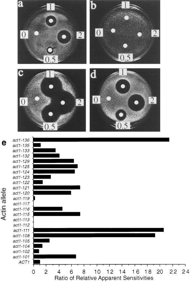
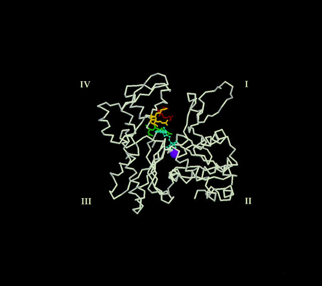
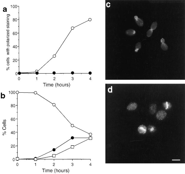
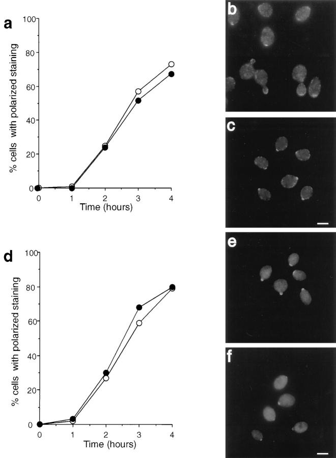
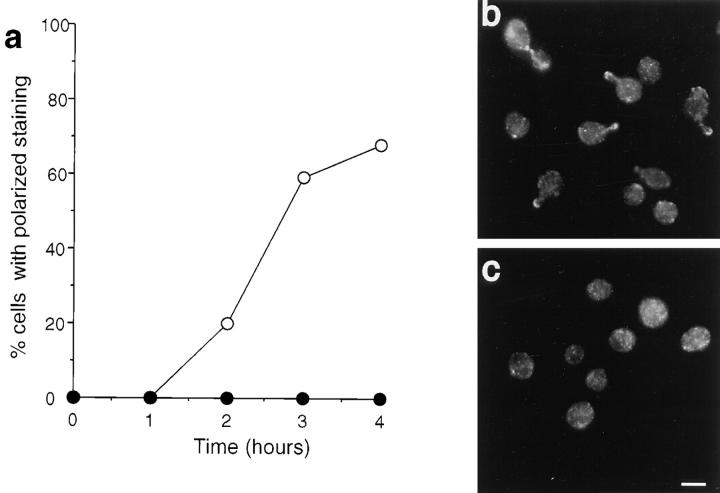
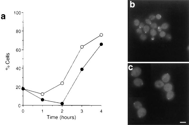
We report that the actin assembly inhibitor latrunculin-A (LAT-A) causes complete disruption of the yeast actin cytoskeleton within 2-5 min, suggesting that although yeast are nonmotile, their actin filaments undergo rapid cycles of assembly and disassembly in vivo. Differences in the LAT-A sensitivities of strains carrying mutations in components of the actin cytoskeleton suggest that tropomyosin, fimbrin, capping protein, Sla2p, and Srv2p act to increase actin cytoskeleton stability, while End3p and Sla1p act to decrease stability. Identification of three LAT-A resistant actin mutants demonstrated that in vivo effects of LAT-A are due specifically to impairment of actin function and implicated a region on the three-dimensional actin structure as the LAT-A binding site. LAT-A was used to determine which of 19 different proteins implicated in cell polarity development require actin to achieve polarized localization. Results show that at least two molecular pathways, one actin-dependent and the other actin-independent, underlie polarity development. The actin-dependent pathway localizes secretory vesicles and a putative vesicle docking complex to sites of cell surface growth, providing an explanation for the dependence of polarized cell surface growth on actin function. Unexpectedly, several proteins that function with actin during cell polarity development, including an unconventional myosin (Myo2p), calmodulin, and an actin-interacting protein (Bud6/Aip3p), achieved polarized localization by an actin-independent pathway, revealing interdependence among cell polarity pathways. Finally, transient actin depolymerization caused many cells to abandon one bud site or mating projection and to initiate growth at a second site. Thus, actin filaments are also required for maintenance of an axis of cell polarity.


Figures

Figure 1
Figure 1
The effect of LAT-A on actin structures in yeast cells. Rd-phalloidin was used to visualize the effects of LAT-A addition to yeast cells. (a) Cells incubated in the absence of LAT-A. (b) Cells incubated for 2 h in the presence of 200 μM LAT-A. The kinetics of the effect of LAT-A on actin structures in yeast cells. (c) Cells were fixed at various time points after the addition of LAT-A. The cells were then processed for visualization of actin using Rd-phalloidin. (d) After 15 min of treatment with LAT-A, the cells were washed twice with fresh medium, resuspended in medium, and allowed to resume growth. Cells were fixed at various time points after washing and processed as above. (▴) actin cables, (○) depolarized cortical actin patches, and (•) polarized actin patches. In both c and d, in each of three repeated experiments, 500 cells were analyzed for their actin structures at each time point. Bar (b) 5 μm.
Figure 2
Figure 2
Sensitivity of a set of congenic actin mutants to LAT-A. Halo assays were used to assess the sensitivity of 23 actin mutants to LAT-A. (a–d) Representative examples of LAT-A halo assays. Concentrations measured 0.5 mM, 1 mM, and 2 mM. (a) ACT1 (wild-type actin), (b) act1-117, (c) act1-129, (d) act1-116. (e) Summary bar graph of the relative apparent sensitivities of all of the actin alleles tested compared to the wild-type.
Figure 3
Figure 3
Mapping the three LAT-A resistant alleles on the actin molecular structure. Backbone of the actin monomer from the coordinates of rabbit muscle actin as determined by Kabsch et al. (1990). Subdomains are marked I to IV. The side chains of the residues mutated to alanine in the LAT-A resistant mutants are shown and are color coded by their allele designation (Wertman et al., 1992). act1112 (yellow), act1-113 (red), act1-117 (green). The adenine nucleotide (cyan) is shown in the prominent cleft as a ball and stick model, and the divalent cation (purple) as a Van der Waal's sphere.
Figure 4
Figure 4
The effect of LAT-A on actin nucleotide exchange. Purified wild-type yeast actin was added to buffer containing a fluorescent nucleotide analogue (ε-ATP) and increasing concentrations of LAT-A. The kinetics of nucleotide exchange were monitored using ε-ATP fluorescence as described in the Materials and Methods section.
Figure 5
Figure 5
The effect of LAT-A on actin polymerization. Purified actin from ACT1 or act1-117 cells was incubated with LAT-A before addition of polymerization salts. (a) After polymerization actin was spun to pellet filamentous actin. Supernatants and pellets were run on gels and actin was visualized using Coomassie staining. (b) Quantitation of the data in a using densitometry. (c) After polymerization, Rd-phalloidin–labeled actin filaments were visualized using fluorescence microscopy. Bar, 5 μM.
Figure 6
Figure 6
The effect of LAT-A on growth and viability of cells. Growth of wild-type cells in the presence (•) or absence (○) of LAT-A assessed by cell counting (a) or by OD measurements (b). Growth of mutant cells in the presence (•) or absence (○) of LAT-A assessed by cell counting, (c) act1-117, and (d) act1-113. (e) Viability of wild-type cells following addition of 100 μM LAT-A to a log phase culture.
Figure 7
Figure 7
Immunolocalization of actin in cells exiting G0 at 25°C in the absence or presence of LAT-A. (a) Quantification of immunofluorescence to assess the percentage of cells incubated in the presence (•) or absence (○) of LAT-A that exhibited polarized actin staining. (b) Morphology of cells exiting G0 in the absence of LAT-A. Cells were classified as unbudded (○), small-budded (•), or medium- to large-budded (□). (c) Immunolocalization of actin in cells grown for 4 h in the absence of LAT-A. (d) Immunolocalization of actin in cells grown for 4 h in the presence of LAT-A. Note the presence in some cells of actin bars and actin in the nucleus (as confirmed by DAPI staining, not shown). Bar, 5 μm.
Figure 8
Figure 8
Immunolocalization of polarity establishment proteins in cells exiting stationary phase. Quantification of immunofluorescence to assess the percentage of cells incubated in the presence (•) or absence (○) of LAT-A that exhibited polarized Cdc42p staining (a) or Bem1p staining (d). Immunolocalization of Cdc42p (b) or Bem1p (e) in cells grown in the absence of LAT-A. Immunolocalization of Cdc42p (c) or Bem1p (f) in cells grown in the presence of LAT-A. Bar, 5 μm.
Figure 13
Figure 13
Polarization of presumptive bud site proteins in the absence (▒⃞ ) or presence (▪) of LAT-A. A summary of data collected for all the proteins analyzed in this study. The cell counts shown here are those at the 4-h time point after release of cells from the stationary phase.
Figure 9
Figure 9
Immunolocalization of Sec4p in cells exiting stationary phase in the absence or presence of LAT-A. (a) Quantification of immunofluorescence to assess the percentage of cells incubated in the presence (•) or absence (○) of LAT-A that exhibited polarized Sec4p staining. Immunolocalization of Sec4p in cells grown in the absence (b) or presence (c) of LAT-A. Bar, 5 μm.
Figure 10
Figure 10
Immunolocalization of calmodulin in cells exiting stationary phase in the absence or presence of LAT-A. (a) Quantification of immunofluorescence to assess the percentage of cells that exhibited polarized calmodulin staining when incubated in the presence (•) or absence (○) of LAT-A. Immunolocalization of calmodulin in cells grown in the absence (b) or presence (c) of LAT-A. Bar, 5 μm.
Figure 11
Figure 11
Immunolocalization of Cdc11p in cells exiting stationary phase in the absence or presence of LAT-A. (c) Quantification of immunofluorescence to assess the percentage of cells that exhibited polarized Cdc11p staining when incubated in the presence (•) or absence (○) of LAT-A. Immunolocalization of Cdc11p in cells grown in the absence (b) or presence (c) of LAT-A. Note the lack of double ring structures in the cells incubated with LAT-A. Bar, 5 μm.
Figure 12
Figure 12
Immunolocalization of Spa2p in cells exiting stationary phase in the absence or presence of LAT-A. (a) Quantification of immunofluorescence to assess the percentage of cells that exhibited polarized Spa2p staining when incubated in the presence (•) or absence (○) of LAT-A. Immunolocalization of Spa2p in cells grown in the absence (b) or presence (c) of LAT-A. Bar, 5 μm.
Figure 14
Figure 14
The formation of two-budded cells after incubation of wild-type cells with LAT-A. Cells were released from stationary phase and allowed to grow until more than 50% of the population was small budded. Cells were then treated with LAT-A for 5 min (or with an equal volume of DMSO in the control case). They were then washed to remove LAT-A and allowed to resume growth. After 2 1/2 h the morphology of cells was assessed. Examples of two-budded haploid cells (a) and diploid cells (c) are shown. Cells were classified as unbudded, single-budded, or two-budded in the haploid (b) or diploid (d) populations. Control cells (▪); cells treated with LAT-A (▨ ).
Figure 15
Figure 15
Model depicting the development of polarity at the presumptive bud site. Cdc42p and Bem1p localize to the presumptive bud site in an actin-independent manner. After localization of these polarity establishment proteins, other proteins associated with the development of cell polarity are able to localize. Proteins associated with secretion require actin in order to achieve their polarized localization. Other proteins do not require actin for localization and therefore occupy a separate branch in polarity development. Note that in the case of Cdc10p, Cdc11p, and Spa2p, a pathway parallel to the actin/secretion pathway is indicated by results from previous studies (see text). Additional experiments are required to elucidate dependency relationships for localization of Cdc10p, Cdc11p, Spa2p, Bni4p, and Gin4p.

References

    1. Adams AEM, Pringle JR. Relationship to actin and tubulin distribution to bud growth in wild type and morphogenetic mutant Saccharomyces cerevisiae. . J Cell Biol. 1984;98:934–945. - PMC - PubMed
    1. Adams AE, Johnson DI, Longnecker RM, Sloat BF, Pringle JR. CDC42 and CDC43 , two additional genes involved in budding and the establishment of cell polarity in the yeast Saccharomyces cerevisiae. . J Cell Biol. 1990;111:131–142. - PMC - PubMed
    1. Amberg DC, Basart E, Botstein D. Defining protein interactions with yeast actin in vivo. Nature Structural Biology. 1995;2:28–35. - PubMed
    1. Amberg, D.C., J.E. Zahner, J.W. Mulholland, J.R. Pringle, and D. Botstein. 1997. Aip3p/Bud6p, a yeast actin-interacting protein that is involved in morphogenesis and the selection of bipolar budding sites. Mol. Biol. Cell. In press. - PMC - PubMed
    1. Ayscough, K.R., and D.G. Drubin. 1997. Immunofluorescence microscopy of yeast cells. In Cell Biology: A Laboratory Handbook. 2nd Edition. J.E. Celis, Editor. Academic Press, San Diego, CA. In press.

Publication types

MeSH terms