. 1997 Apr 21;137(2):417-31.
doi: 10.1083/jcb.137.2.417.

The Saccharomyces cerevisiae kinesin-related motor Kar3p acts at preanaphase spindle poles to limit the number and length of cytoplasmic microtubules

Affiliations

The Saccharomyces cerevisiae kinesin-related motor Kar3p acts at preanaphase spindle poles to limit the number and length of cytoplasmic microtubules

W Saunders et al. J Cell Biol. .

Abstract

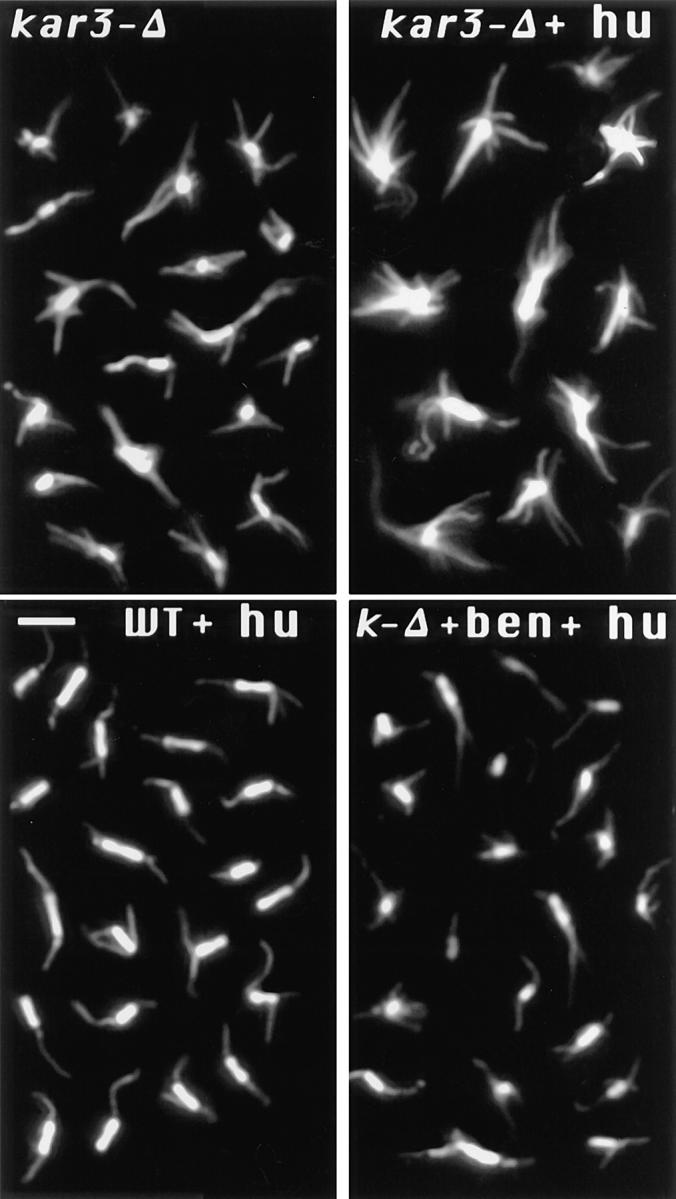
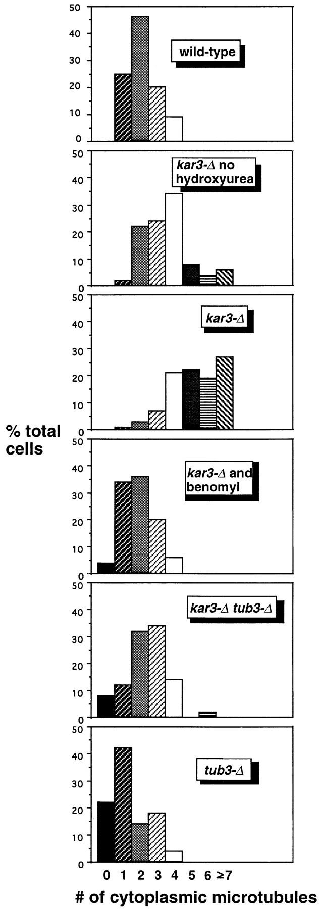
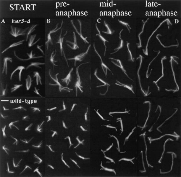
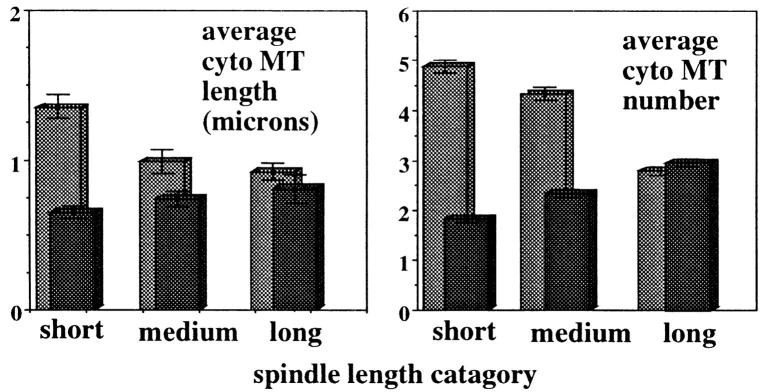
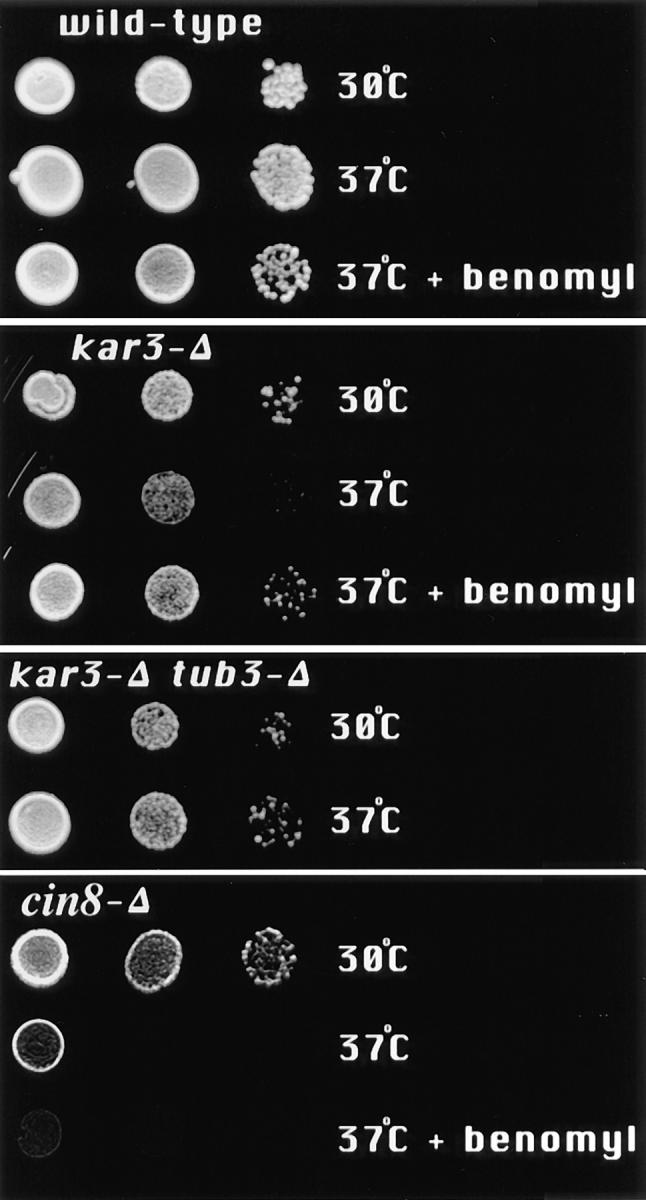
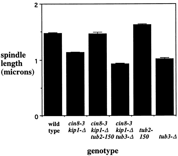
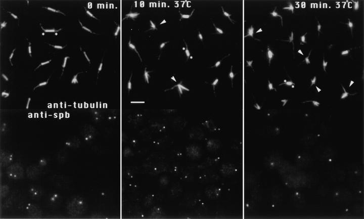
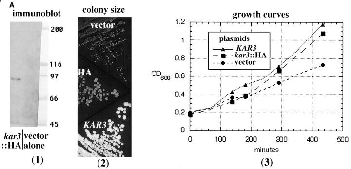
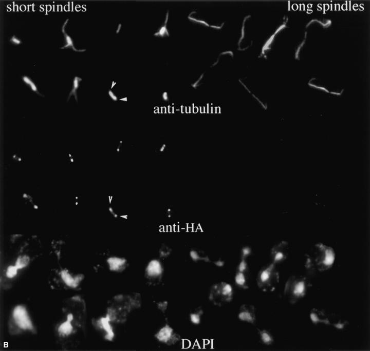
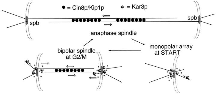
The Saccharomyces cerevisiae kinesin-related motor Kar3p, though known to be required for karyogamy, plays a poorly defined, nonessential role during vegetative growth. We have found evidence suggesting that Kar3p functions to limit the number and length of cytoplasmic microtubules in a cell cycle-specific manner. Deletion of KAR3 leads to a dramatic increase in cytoplasmic microtubules, a phenotype which is most pronounced from START through the onset of anaphase but less so during late anaphase in synchronized cultures. We have immunolocalized HA-tagged Kar3p to the spindle pole body region, and fittingly, Kar3p was not detected by late anaphase. A microtubule depolymerizing activity may be the major vegetative role for Kar3p. Addition of the microtubule polymerization inhibitors nocodazol or benomyl to the medium or deletion of the nonessential alpha-tubulin TUB3 gene can mostly correct the abnormal microtubule arrays and other growth defects of kar3 mutants, suggesting that these phenotypes result from excessive microtubule polymerization. Microtubule depolymerization may also be the mechanism by which Kar3p acts in opposition to the anaphase B motors Cin8p and Kip1p. A preanaphase spindle collapse phenotype of cin8 kip1 mutants, previously shown to involve Kar3p, is markedly delayed when microtubule depolymerization is inhibited by the tub2-150 mutation. These results suggest that the Kar3p motor may act to regulate the length and number of microtubules in the preanaphase spindle.


Figures

Figure 1
Figure 1
Change in spindle structure with loss of Kar3p. kar3-Δ or wild-type (WT) cells were arrested with hydroxyurea (hu) or grown to log phase without arrest, and samples were fixed and processed for anti-tubulin immunofluorescence. Each panel is a composite image of representative preanaphase spindles from a single sample. As shown, loss of KAR3 led to an increase in cytoplasmic microtubules in unarrested cultures (kar3-Δ). This phenotype was greatly exaggerated after mitotic arrest with hydroxyurea (kar3-Δ + hu). Short spindles in wild-type cells appeared essentially the same with or without hydroxyurea treatment (shown with hydroxyurea treatment). Addition of 10 μg/ml benomyl during the hydroxyurea arrest prevented the abnormal spindle structure in kar3 mutants. Bar, 2 μm.
Figure 2
Figure 2
The increased cytoplasmic microtubule number in hydroxyurea-arrested kar3-Δ cells can be corrected by addition of benomyl or deletion of TUB3. All samples were arrested with hydroxyurea (except as indicated) for 4 h at 26°C, fixed, and stained with anti-tubulin antibodies by indirect immunofluorescence (see Materials and Methods). Cells were examined by epifluorescence microscopy, and the approximate number of cytoplasmic microtubules was determined. Cytoplasmic microtubules were defined as those that were too long to be nuclear and/or that clearly pointed away from the nuclear envelope. Loss of Kar3p resulted in an increase in cytoplasmic microtubules, especially in the hydroxyurea-arrested culture. This defect could be mostly corrected by addition of 10 μg/ ml benomyl to the culture medium or by deletion of the TUB3 genomic locus. tub3-Δ single mutants had fewer cytoplasmic microtubules than wild-type cells, while the tub3-Δ kar3-Δ double mutants had a number intermediate between that of tub3-Δ and kar3-Δ single mutants. The number of cytoplasmic microtubules from 100 cells was determined for each sample. Note that these numbers may represent an underestimate of the total, as some short microtubules (∼25% of the total) could not be determined to be nuclear or cytoplasmic. Also, the most abnormal spindles in the kar3-Δ populations were probably not recognized as spindles and not included in the sample.
Figure 3
Figure 3
Cytoplasmic microtubule numbers in α-factor–arrested and released kar3-Δ mutants. Wild-type and mutant cells were arrested with α-factor, washed, and released in fresh medium. Samples were fixed and processed for anti-tubulin immunofluorescence for 0 (A) and 90 min (B–D) of release. α-Factor–arrested kar3 mutants had more and longer microtubules than arrested wild-type cells. For the 90 min release samples, selected spindles are shown from the same sample which are short (B), medium (C), or longer in length (D). Short spindles most likely represent cells that have not yet begun or are in the earliest stages of anaphase. kar3 mutant cells at this stage have markedly abnormal microtubule arrays, similar to those seen in the hydroxyurea-arrested cultures (compare to Fig. 1). Medium length spindles are typically from cells that have started but not completed anaphase, and in the kar3 mutants are closer in appearance to those from wild-type cells. Cells with the longest spindles are presumed to be in late anaphase or early telophase. These spindles are almost indistinguishable between wild-type cells and kar3 mutants. Bar, 2 μm.
Figure 4
Figure 4
Measurements of cytoplasmic microtubule numbers and lengths in α-factor–arrested and released cells. Wild-type and kar3 mutants were arrested and released as in Fig. 3. The numbers and lengths of the cytoplasmic microtubules were counted as in Fig. 2. For this analysis, short spindles were defined as those where no chromatin separation was visible (typically 2.5 μm or less). Medium length spindles were those with spindles between ∼2.5 and 7.0 μm in length and at least some chromatin separation visible. Longer spindles were those greater than ∼7.0 μm in length with well separated chromatin masses. 100 spindles were examined for each category. As shown, the number and length of cytoplasmic microtubules in wild-type cells (black) increased slightly with spindle length. kar3 mutants (gray) with short spindles had many more and longer cytoplasmic microtubules than wild-type cells. Late anaphase spindles had similar numbers of cytoplasmic microtubules in wild-type and kar3 mutants.
Figure 5
Figure 5
Temperature sensitivity of strains containing the kar3-Δ allele was mostly corrected by addition of benomyl to the medium or deletion of TUB3. Cells of the indicated genotypes were suspended in water and serial dilutions placed on YPD plates with or without 5 μg/ml benomyl, at 30° or 37°C, for 2–3 d. Growth of kar3-Δ cells was observed to be partially inhibited at 37°C. This temperature sensitivity was eliminated by addition of benomyl to the medium or by deletion of TUB3. The benomyl sensitivity caused by the tub3-Δ allele was not rescued by kar3-Δ (not shown). The kar3-Δ, tub3-Δ, and tub3-Δ kar3-Δ double mutants are all from the same tetrad. (Growth of kar3-Δ mutants at 37°C did not cause a noticeable cell cycle arrest phenotype, results not shown.) In contrast, deletion of CIN8, which also causes slight temperature sensitivity, could not be rescued by benomyl.
Figure 6
Figure 6
The short spindle phenotype from cin8 kip1 mutations is sensitive to tubulin mutations. Cells with the indicated genotypes were arrested with hydroxyurea at 26°C (a permissive temperature for growth of these strains), fixed, stained with antitubulin antibody, and the length of the spindles measured (see Materials and Methods). Mutational inhibition of the Cin8p and Kip1p motors produced a decrease in spindle length. This defect could be corrected by the tub2-150 mutation and was worsened by the tub3-Δ mutation. The numbers given represent the combined averages for two separate strains containing the indicated allele(s). Complete data for the individual strains are given in Table I. Each value represents at least 200 spindles counted during two to four separate experiments.
Figure 7
Figure 7
The tub2-150 allele slowed down the spindle collapse in cin8 kip1 mutants. cin8-3 kip1-Δ tub2-150 mutants were arrested with 0.1 M hydroxyurea for 4 h and transferred to 37°C for 10 and 30 min, still in the presence of hydroxyurea. Samples were fixed and stained with anti-tubulin or anti-spindle pole body antibodies. At 0 min, most cells had intact spindles and duplicated and separated spindle poles. After 10 min at 37°C most of the spindles remained intact but were noticeably shorter than before the temperature shift. After 30 min, most of the spindles had collapsed, and the remaining spindles were shorter still. At this time point only single dots of spindle pole staining were typically observed. The anti-tubulin staining images are composites taken from different fields of the same sample (see Materials and Methods). The anti–spindle pole staining images are not composites and represent single fields. For selected examples, asterisks indicate the position of the spindle poles of intact spindles, and arrowheads point to collapsed spindles. Bar, 2 μm.
Figure 8
Figure 8
Immunolocalization of HA-tagged Kar3p to the spindle poles. A triple HA epitope tag was inserted near the 3′ end of the KAR3 coding sequence (see Materials and Methods) and transformed into a kar3-Δ strain. (A; 1) Immunoblot with anti-HA antibodies. kar3-Δ cells with the HA-tagged KAR3 (pGD15) or with vector alone (Ycp50) were grown in selective medium. Lysed cells were run on an SDS-PAGE gel, blotted to membrane, and probed with anti-HA antibodies (see Materials and Methods). Ponceau S–stained markers from the same gel are also shown. The predicted size of Kar3p is 84 kD. Similar results were seen in multiple experiments. (2) The colony size of kar3 mutants with vector alone (YCp50) was observed to be smaller and more varied in size than strains with KAR3 on a plasmid (pMR798). When HA-tagged KAR3 (pGD15) was transformed into kar3-Δ mutants the plasmid allowed normal colony size. Cells were grown in selective −ura medium overnight, streaked onto the same YEPD plates, and grown at 30°C for 3 d. Different kar3Δ strains gave similar results. (3) Growth curves of kar3 mutants. kar3-Δ cells were observed to double at similar rates with plasmids containing KAR3 (▴) or HA-tagged kar3 (▪), while those containing vector alone (•) divided more slowly. This test was repeated twice with similar results. (B) Cells grown to log phase were fixed briefly with formaldehyde and subjected to triple staining with antibodies to HA and tubulin and with DAPI. As shown, most of the HA-tagged Kar3p was observed to be associated with the spindle poles. The spindle pole bodies in a sample spindle are marked with arrowheads. Some intrapolar staining could be seen. Staining of intermediate length spindles varied (not shown), but long spindles from anaphase cells were invariably negative. Images shown are a composite of cells from the same sample.
Figure 8
Figure 8
Immunolocalization of HA-tagged Kar3p to the spindle poles. A triple HA epitope tag was inserted near the 3′ end of the KAR3 coding sequence (see Materials and Methods) and transformed into a kar3-Δ strain. (A; 1) Immunoblot with anti-HA antibodies. kar3-Δ cells with the HA-tagged KAR3 (pGD15) or with vector alone (Ycp50) were grown in selective medium. Lysed cells were run on an SDS-PAGE gel, blotted to membrane, and probed with anti-HA antibodies (see Materials and Methods). Ponceau S–stained markers from the same gel are also shown. The predicted size of Kar3p is 84 kD. Similar results were seen in multiple experiments. (2) The colony size of kar3 mutants with vector alone (YCp50) was observed to be smaller and more varied in size than strains with KAR3 on a plasmid (pMR798). When HA-tagged KAR3 (pGD15) was transformed into kar3-Δ mutants the plasmid allowed normal colony size. Cells were grown in selective −ura medium overnight, streaked onto the same YEPD plates, and grown at 30°C for 3 d. Different kar3Δ strains gave similar results. (3) Growth curves of kar3 mutants. kar3-Δ cells were observed to double at similar rates with plasmids containing KAR3 (▴) or HA-tagged kar3 (▪), while those containing vector alone (•) divided more slowly. This test was repeated twice with similar results. (B) Cells grown to log phase were fixed briefly with formaldehyde and subjected to triple staining with antibodies to HA and tubulin and with DAPI. As shown, most of the HA-tagged Kar3p was observed to be associated with the spindle poles. The spindle pole bodies in a sample spindle are marked with arrowheads. Some intrapolar staining could be seen. Staining of intermediate length spindles varied (not shown), but long spindles from anaphase cells were invariably negative. Images shown are a composite of cells from the same sample.
Figure 9
Figure 9
Model for Kar3p function in microtubule arrays. The Kar3p motor (shown as shaded circles) is proposed to function predominantly at spindle poles to stimulate microtubule turnover. At START of the cell cycle, the activity of Kar3p is required to limit microtubule number and length at the single spindle pole body (spb). During spindle assembly, the now duplicated spindle poles become linked by microtubule crosslinking and chromosome attachment (latter not shown). Depolymerization of the microtubules linking the spindle poles would now have the effect of pulling the poles together. Cin8p and Kip1p (dark circles) apparently function to counter this inwardly directed force, most likely by crosslinking and sliding the nuclear microtubules, using the microtubules to push out on the spindle poles (Hoyt et al., 1992) though other mechanisms are also possible. The bipolar spindle now has two types of forces acting in opposition on the spindle poles, represented by open arrows. Once anaphase begins, the antagonistic relationship between Cin8p and Kar3p changes. Kar3p-driven spindle collapse no longer occurs in cin8 kip1 mutants (Saunders et al., 1992); Kar3p is no longer found at the spindle poles, and there is a net polymerization of the spindle microtubules (and to a lesser extent the cytoplasmic microtubules) resulting in a great increase in spindle length. Thus the outwardly directed force of Cin8p and Kip1p are retained, but the inward pull of Kar3p is lost. It is proposed that this transition is a major contributor to the onset of anaphase B and can occur in the absence of chromosomes (Zhang and Nicklas, 1996). Spindle pole bodies are shown as discs embedded in a nuclear envelope, small open circles represent microtubule depolymerization, and dark arrows the presumed direction of Kar3p movement.

References

    1. Belmont LD, Hyman AA, Sawin KE, Mitchison TJ. Real-time visualization of cell cycle dependent changes in microtubule dynamics in cytoplasmic extracts. Cell. 1990;62:579–589. - PubMed
    1. Buendia B, Draetta G, Karsenti E. Regulation of the microtubule nucleating activity of centrosomes in Xenopusegg extracts: role of cyclin A–associated protein kinase. J Cell Biol. 1992;116:1431–1442. - PMC - PubMed
    1. Davidse LC. Benzimadazole fungicides: mechanism of action and biological impact. Annu Rev Phytopathol. 1986;24:43–65.
    1. De Brabander M. Microtubule dynamics during the cell cycle: the effects of taxol and nocodazole on the microtubule system of PtK2 cells at different stages of the mitotic cycle. Int Rev Cytol. 1986;101:215–274. - PubMed
    1. Desai A, Mitchison T. A new role for motor proteins as couplers to depolymerizing microtubules. J Cell Biol. 1995;128:1–4. - PMC - PubMed

Publication types

MeSH terms