. 1997 Apr 21;137(2):445-58.
doi: 10.1083/jcb.137.2.445.

Synaptic-like microvesicles of neuroendocrine cells originate from a novel compartment that is continuous with the plasma membrane and devoid of transferrin receptor

Affiliations

Synaptic-like microvesicles of neuroendocrine cells originate from a novel compartment that is continuous with the plasma membrane and devoid of transferrin receptor

A Schmidt et al. J Cell Biol. .

Erratum in

  • J Cell Biol 1997 Jun 2;137(5):1197

Abstract

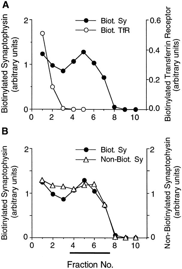
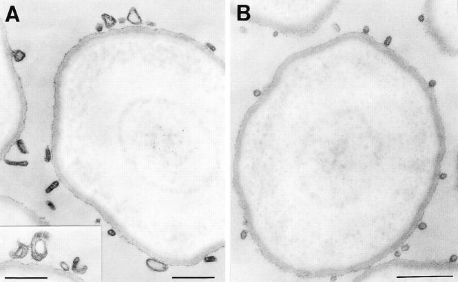
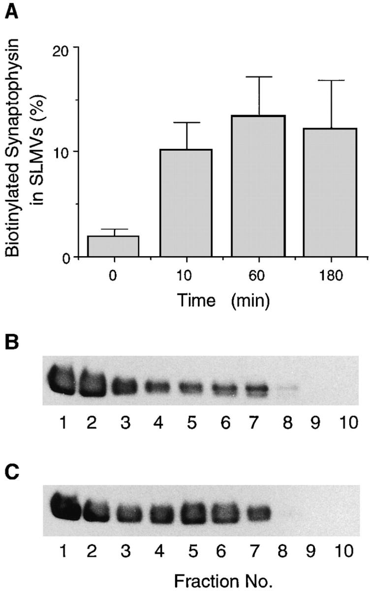
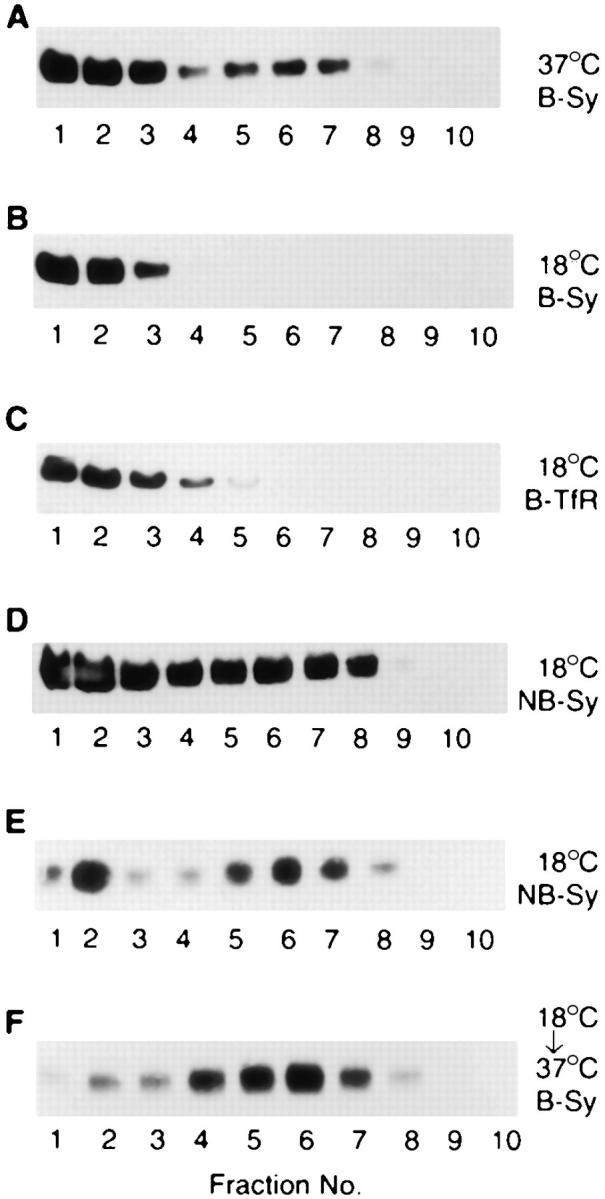
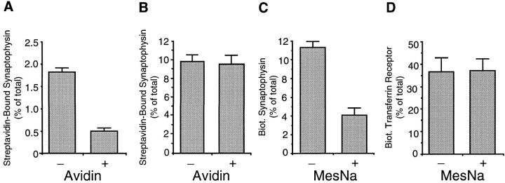
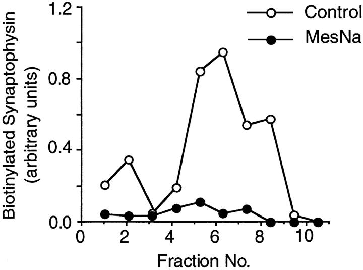
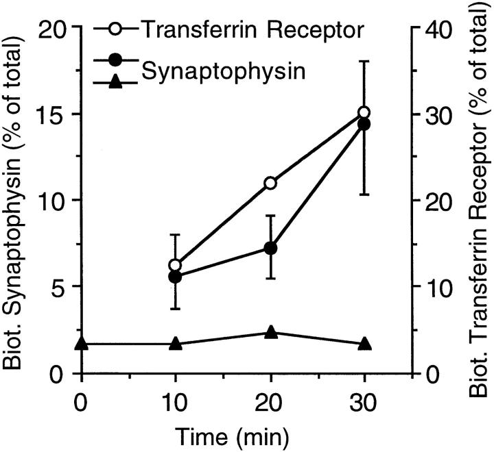
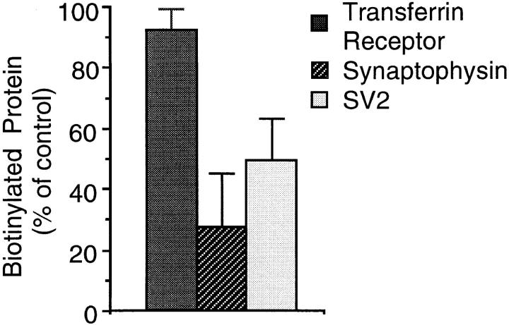
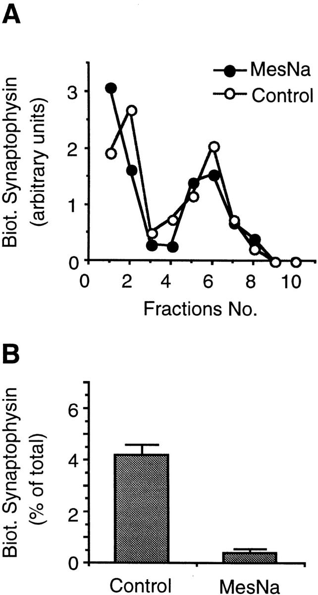
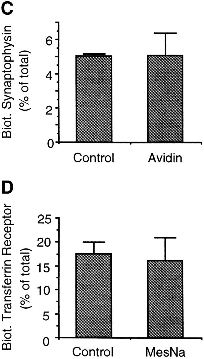
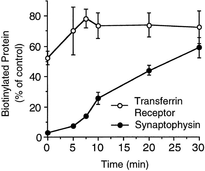
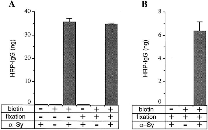
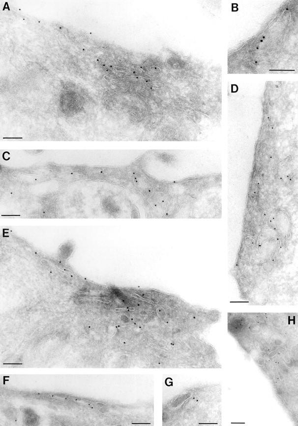
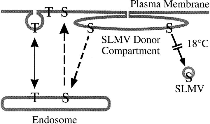
We have characterized the compartment from which synaptic-like microvesicles (SLMVs), the neuroendocrine counterpart of neuronal synaptic vesicles, originate. For this purpose we have exploited the previous observation that newly synthesized synaptophysin, a membrane marker of synaptic vesicles and SLMVs, is delivered to the latter organelles via the plasma membrane and an internal compartment. Specifically, synaptophysin was labeled by cell surface biotinylation of unstimulated PC12 cells at 18 degrees C, a condition which blocked the appearance of biotinylated synaptophysin in SLMVs and in which there appeared to be no significant exocytosis of SLMVs. The majority of synaptophysin labeled at 18 degrees C with the membrane-impermeant, cleavable sulfo-NHS-SS-biotin was still accessible to extracellularly added MesNa, a 150-D membrane-impermeant thiol-reducing agent, but not to the 68,000-D protein avidin. The SLMVs generated upon reversal of the temperature to 37 degrees C originated exclusively from the membranes containing the MesNa-accessible rather than the MesNa-protected population of synaptophysin molecules. Biogenesis of SLMVs from MesNa-accessible membranes was also observed after a short (2 min) biotinylation of synaptophysin at 37 degrees C followed by chase. In contrast to synaptophysin, transferrin receptor biotinylated at 18 degrees or 37 degrees C became rapidly inaccessible to MesNa. Immunofluorescence and immunogold electron microscopy of PC12 cells revealed, in addition to the previously described perinuclear endosome in which synaptophysin and transferrin receptor are colocalized, a sub-plasmalemmal tubulocisternal membrane system distinct from caveolin-positive caveolae that contained synaptophysin but little, if any, transferrin receptor. The latter synaptophysin was selectively visualized upon digitonin permeabilization and quantitatively extracted, despite paraformaldehyde fixation, by Triton X-100. Synaptophysin biotinylated at 18 degrees C was present in these subplasmalemmal membranes. We conclude that SLMVs originate from a novel compartment that is connected to the plasma membrane via a narrow membrane continuity and lacks transferrin receptor.


Figures

Figure 1
Figure 1
Biotinylated synaptophysin is sorted to SLMVs. PC12 cells were incubated with sulfo-NHS-LC–biotin for 5 min at 37°C and chased for 180 min at 37°C. The 12,000-g supernatant prepared from the cells was subjected to glycerol gradient centrifugation, and the fractions (n 1, bottom) were analyzed for biotinylated (A and B, closed circles, same data points for both panels) and nonbiotinylated (B, open triangles) synaptophysin and biotinylated transferrin receptor (A, open circles) by streptavidin–agarose adsorption followed by immunoblotting of bound and unbound material with the respective antibodies. Bar, SLMVs.
Figure 2
Figure 2
Electron microscopy of glycerol gradient fractions. The 12,000 g supernatant prepared from PC12 cells was subjected to glycerol gradient centrifugation, and the pools of fractions 1 and 2 (A) and fractions 5–9 (B) were subjected to immunoisolation using anti-synaptophysin beads followed by fixation and processing for electron microscopy. Note the heterogeneous membrane structures in A and the homogenous population of 50-nm vesicles in B. No membranes were adsorbed to the beads if the anti-synaptophysin antibody was omitted (not shown). Bars, 300 nm.
Figure 3
Figure 3
Time course of appearance of biotinylated synaptophysin in SLMVs. PC12 cells were incubated with sulfo-NHS-LC–biotin for 5 min at 37°C and chased at 37°C for various times as indicated (A) or for 3 min (B) and 180 min (C). The 66,000 g (A) or 12,000 g (B and C) supernatants prepared from the cells were subjected to glycerol gradient centrifugation, and the fractions were analyzed for biotinylated synaptophysin by streptavidin–agarose adsorption followed by immunoblotting of bound material with antisynaptophysin. (A) Synaptophysin immunoreactivity in the SLMV-containing fractions is expressed as percent of total biotinylated synaptophysin. Data are the mean of two (0, 10, 180 min) or three (60 min) independent experiments; bars indicate the variation of individual values from the mean or the standard error, respectively. (B and C) Immunoblots; 2.5% (B) and 11% (C) of the total biotinylated synaptophysin was recovered in the SLMV-containing fractions (B, n 5–8; C, n 4–8).
Figure 4
Figure 4
Biotinylated synaptophysin does not appear in SLMVs at 18°C. PC12 cells were incubated with sulfoNHS-LC–biotin either for 30 min at 37°C (A), for 30 min at 18°C (B–E), or for 30 min at 18°C followed by a 30-min chase at 37°C (F). The 12,000 g (A–D) or 66,000 g (E and F) supernatants prepared from the cells were subjected to glycerol gradient centrifugation, and the fractions were analyzed for biotinylated (BSy; A, B, and F) and nonbiotinylated (NB-Sy; D and E) synaptophysin and biotinylated transferrin receptor (BTfR; C) by streptavidin–agarose adsorption followed by immunoblotting of bound (A–C and F) and unbound (D and E) material with the respective antibodies. The immunoblots shown in (B) and (C) were obtained from the same filter. 16% (A) and 15% (F) of the total biotinylated synaptophysin was recovered in the SLMV-containing fractions (A, n 5–8; F, n 4–8); the immunoblot shown in F is a longer exposure relative to that shown in A. Note that the ratio of SLMVs to the larger membranes recovered in the bottom fractions of the gradient is greater in E than D, because a 66,000 g and a 12,000 g supernatant, respectively, was subjected to glycerol gradient centrifugation.
Figure 5
Figure 5
Differential sensitivity to extracellular probes of synaptophysin and transferrin receptor biotinylated at 18°C. PC12 cells were incubated with sulfo-NHS-LC– biotin (A and B) or sulfoNHS-SS–biotin (C and D) for 60 min at 4°C (A) or for 30 min at 18°C (B–D), chased for 5 min at 18°C in the presence of glycine (B–D) or not chased (A), and incubated at 4°C in the absence (−) or presence (+) of extracellularly added avidin (A and B) or MesNa (C and D). Synaptophysin and transferrin receptor in the postnuclear supernatants were analyzed for binding to streptavidin–agarose by immunoblotting of bound and unbound material with the respective antibodies. Streptavidin-bound biotinylated synaptophysin and transferrin receptor present in the postnuclear supernatant is expressed as percentage of total (sum of streptavidin-bound and streptavidin-unbound synaptophysin and transferrin receptor, respectively). (A) Data are the mean of two independent experiments; bars indicate the variation of the individual values from the mean. (B–D) Data are the mean of three independent experiments; bars indicate SD.
Figure 7
Figure 7
SLMV biogenesis involves the MesNa-sensitive population of synaptophysin molecules biotinylated at 18°C. PC12 cells were incubated with sulfo-NHS-SS–biotin for 30 min at 18°C, chased for 5 min at 18°C in the presence of glycine, incubated at 4°C in the absence (Control) or presence of extracellularly added MesNa, and chased for 10 min at 37°C. The 66,000-g supernatants prepared from the cells were subjected to glycerol gradient centrifugation, and fractions were analyzed for biotinylated synaptophysin by streptavidin– agarose adsorption followed by immunoblotting of bound material with anti-synaptophysin antibodies. 11% and 4% of the total synaptophysin present in the sum of the 66,000 g pellet plus supernatant bound to streptavidin–agarose without and with MesNa addition before the 37°C chase, respectively.
Figure 6
Figure 6
Time course of accumulation of biotinylated synaptophysin and transferrin receptor at 18°C. PC12 cells were incubated with (open and closed circles) or without (closed triangles) sulfo-NHS-LC–biotin at 18°C for the indicated times. The cells incubated without sulfoNHS-LC–biotin at 18°C were subsequently biotinylated for 30 min at 4°C (closed triangles). Postnuclear supernatants prepared from the cells were analyzed for biotinylated and nonbiotinylated synaptophysin and transferrin receptor by streptavidin– agarose adsorption, followed by immunoblotting of bound and unbound material with the respective antibodies. Biotinylated synaptophysin and transferrin receptor is expressed as percent of total (sum of biotinylated plus nonbiotinylated synaptophysin and transferrin receptor, respectively). Data points without error bars represent single determinations. Data points with error bars represent the mean of three (transferrin receptor), four (synaptophysin at 10 and 30 min), or two (synaptophysin at 20 min) independent determinations; bars indicate SD or the variation of the individual values from the mean.
Figure 8
Figure 8
SV2 is accessible to MesNa after biotinylation at 18°C. PC12 cells were incubated with sulfo-NHS-SS– biotin for 30 min at 18°C, chased for 5 min at 18°C in the presence of glycine, and incubated at 4°C in the absence (control) or presence of extracellularly added MesNa. Transferrin receptor, synaptophysin, and SV2 in the postnuclear supernatants were analyzed for binding to streptavidin–agarose by immunoblotting of bound and unbound material with the respective antibodies. Streptavidin-bound biotinylated transferrin receptor, synaptophysin, and SV2 present in the postnuclear supernatant were calculated as percentage of total (sum of streptavidin-bound plus streptavidinunbound transferrin receptor, synaptophysin, and SV2, respectively), and the individual values obtained after MesNa treatment were expressed as percentage of control. Data are the mean of three independent experiments; bars indicate SD. The mean values of biotinylated protein for the control condition were 25.3% (transferrin receptor), 7.5% (synaptophysin), and 3.1% (SV2).
Figure 9
Figure 9
SLMVs originate from an avidin-protected, MesNa-accessible compartment at 37°C. PC12 cells were incubated with sulfoNHS-SS–biotin for 2 min at 37°C, chased for 10 min at 37°C, and incubated at 4°C in the absence (Control) or presence of extracellularly added MesNa (A, B, and D) or avidin (C). The 66,000 g pellets and supernatants were prepared from the cells and the supernatants subjected to glycerol gradient centrifugation. Synaptophysin in the glycerol gradient fractions (A) and transferrin receptor and synaptophysin in the 66,000 g pellets (B–D) were analyzed for binding to streptavidin– agarose by immunoblotting of bound and unbound material with the respective antibodies. (A) A representative experiment showing biotinylated synaptophysin in the glycerol gradient fractions. (B–D) Biotinylated synaptophysin (B and C) and transferrin receptor (D) in the 66,000 g pellet is expressed as percent of total (sum of streptavidin-bound plus streptavidin-unbound synaptophysin and transferrin receptor, respectively, present in the sum of 66,000 g pellet plus supernatant). Data are the mean of four (MesNa) or two (Avidin) independent experiments; bars indicate SD or the variation of the individual values from the mean.
Figure 9
Figure 9
SLMVs originate from an avidin-protected, MesNa-accessible compartment at 37°C. PC12 cells were incubated with sulfoNHS-SS–biotin for 2 min at 37°C, chased for 10 min at 37°C, and incubated at 4°C in the absence (Control) or presence of extracellularly added MesNa (A, B, and D) or avidin (C). The 66,000 g pellets and supernatants were prepared from the cells and the supernatants subjected to glycerol gradient centrifugation. Synaptophysin in the glycerol gradient fractions (A) and transferrin receptor and synaptophysin in the 66,000 g pellets (B–D) were analyzed for binding to streptavidin– agarose by immunoblotting of bound and unbound material with the respective antibodies. (A) A representative experiment showing biotinylated synaptophysin in the glycerol gradient fractions. (B–D) Biotinylated synaptophysin (B and C) and transferrin receptor (D) in the 66,000 g pellet is expressed as percent of total (sum of streptavidin-bound plus streptavidin-unbound synaptophysin and transferrin receptor, respectively, present in the sum of 66,000 g pellet plus supernatant). Data are the mean of four (MesNa) or two (Avidin) independent experiments; bars indicate SD or the variation of the individual values from the mean.
Figure 10
Figure 10
Kinetics of the acquisition of MesNa inaccessibility. PC12 cells were incubated with sulfo-NHS-SS– biotin for 2 min at 37°C, chased for the indicated times at 37°C, and incubated at 4°C in the absence (controls, 0 and 30 min chase only) or presence of MesNa. Synaptophysin and transferrin receptor in the cell lysates were analyzed for binding to streptavidin–agarose by immunoblotting of bound and unbound material with the respective antibodies. Synaptophysin and transferrin receptor bound to streptavidin–agarose were calculated as percentage of total (sum of streptavidin-bound plus streptavidinunbound synaptophysin and transferrin receptor, respectively) and are expressed as percentage of control (mean of the control values at 0 and 30 min of chase, which were very similar to each other). Data are the mean of two independent experiments; bars indicate the variation of the individual values from the mean and, for some time points, are within the size of the symbol. In the control, 4.2 ± 0.3% and 11.0 ± 1.3% of the total synaptophysin and transferrin receptor were biotinylated, respectively.
Figure 11
Figure 11
Double fluorescence analysis of PC12 cells for synaptophysin and internalized transferrin. PC12 cells were incubated with Texas red–labeled transferrin for 30 min at 37°C, fixed with paraformaldehyde, permeabilized with either digitonin (A, B, a, and b) or Triton X-100 (C, D, c, and d), and labeled with anti-synaptophysin followed by Cy2- labeled secondary antibody. Cells were analyzed by confocal laser scanning microscopy for synaptophysin immunoreactivity (A, a, C, and c) or internalized transferrin (B, b, D, and d). Single optical sections in the xy (A–D) or xz (a–d) planes of the same cells are shown. The white triangular indentations at the margins of the panels indicate the approximate positions of the respective corresponding plane. The same confocal microscope parameters were used for the image pairs shown in panels A and C, B and D, a and c, and b and d. Note that upon permeabilization with digitonin, synaptophysin immunoreactivity is detected at the cell periphery but not in the perinuclear area which, however, shows transferrin fluorescence. After permeabilization with Triton X-100, the synaptophysin immunoreactivity at the cell periphery is decreased concomitant with its appearance in the perinuclear area, where it is largely colocalized with transferrin.
Figure 12
Figure 12
Synaptophysin biotinylated at 18°C is quantitatively extracted from paraformaldehyde-fixed PC12 cells by Triton X-100 (A) and is accessible to anti-synaptophysin after digitonin permeabilization of fixed cells (B). (A) PC12 cells were incubated without (−) or with (+) sulfo-NHS-LC–biotin for 30 min at 18°C, chased for 5 min at 18°C in the presence of glycine, and fixed (+) or not fixed (−), and Triton X-100 extracts were subjected to streptavidin–agarose adsorption. Specific immunoreactivity due to the binding of biotinylated synaptophysin to streptavidin–agarose was determined by incubating the beads without (−) or with (+) anti-synaptophysin antibody (α-Sy) followed by HRP-conjugated goat anti–mouse IgG antibody. Data are the mean of values obtained from three coverslips; bars indicate SD. (B) PC12 cells were incubated without (−) or with (+) sulfo-NHS-LC– biotin for 30 min at 18°C, chased for 5 min at 18°C in the presence of glycine, fixed, permeabilized with digitonin, incubated without (−) or with (+) anti-synaptophysin (α-Sy), and extracted with Triton X-100. The Triton extracts were subjected to streptavidin– agarose adsorption, and anti-synaptophysin bound to the beads via biotinylated synaptophysin was detected by HRP-conjugated goat anti–mouse IgG antibody. Data are the mean of values obtained from three coverslips; bars indicate SD. The lower synaptophysin immunoreactivity in B than A (compare ordinate scales) presumably reflects incomplete accessibility of the anti-synaptophysin to its epitope when added to digitonin-permeabilized fixed cells as compared with anti-synaptophysin addition after Triton X-100 extraction of synaptophysin and its adsorption to streptavidin–agarose beads.
Figure 13
Figure 13
Morphology of the SLMV donor compartment. PC12 cells incubated at 18° (A–D) or 37°C (E–H) with sulfo-NHS-LC–biotin for 30 min followed by a 5-min chase were fixed at 4°C. Ultrathin cryosections were immunogold-labeled for synaptophysin and analyzed by electron microscopy. Note the synaptophysin immunoreactivity associated with tubulo-cisternal membrane structures beneath the plasma membrane. Little synaptophysin immunoreactivity is found at the plasma membrane itself. Bars, 100 nm.
Figure 14
Figure 14
Model for the biogenesis of SLMVs from a subplasmalemmal compartment connected to the plasma membrane. T, transferrin receptor; S, synaptophysin. Segregation of synaptophysin from the transferrin receptor occurs at the plasma membrane. The transferrin receptor is internalized via endocytic vesicles to endosomes located predominantly in the perinuclear region (MesNa protected), from which it recycles to the plasma membrane. Both endocytosis and recycling occur at 18°C. Synaptophysin, but not the transferrin receptor, moves into the SLMV donor compartment via lateral mobility and/or membrane invagination. The SLMV donor compartment, located at the periphery of the cell, is connected with the plasma membrane via a narrow membrane continuity allowing entry of MesNa, but not avidin, at 4°C. From the SLMV donor compartment, a minor proportion of synaptophysin (10–15%) is incorporated into SLMVs. This process is blocked at 18°C. The majority of synaptophysin is either delivered to perinuclear, MesNa-protected endosomes from which it recycles, like the transferrin receptor, to the plasma membrane (broken arrows) or moves to MesNa-inaccessible membranes connected to the SLMV donor compartment (not illustrated). For further details, see Discussion.

References

    1. Anderson RGW. Potocytosis of small molecules and ions by caveolae. Trends Cell Biol. 1993;3:69–72. - PubMed
    1. Bauerfeind R, Régnier-Vigouroux A, Flatmark T, Huttner WB. Selective storage of acetylcholine, but not catecholamines, in neuroendo-crine synaptic-like microvesicles off early endosomal origin. Neuron. 1993;11:105–121. - PubMed
    1. Baumert M, Maycox PR, Navone F, De Camilli P, Jahn R. Synaptobrevin: an integral membrane protein of 18,000 daltons present in small synaptic vesicles of rat brain. EMBO (Eur Mol Biol Organ) J. 1989;8:379–384. - PMC - PubMed
    1. Bennett MK, Scheller RH. A molecular description of synaptic vesicle membrane trafficking. Annu Rev Biochem. 1994;63:63–100. - PubMed
    1. Buckley K, Kelly RB. Identification of a transmembrane glycoprotein specific for secretory vesicles of neural and endocrine cells. J Cell Biol. 1985;100:1284–1294. - PMC - PubMed

Publication types

MeSH terms