. 1997 Apr 21;137(2):509-19.
doi: 10.1083/jcb.137.2.509.

Isolated P-selectin glycoprotein ligand-1 dynamic adhesion to P- and E-selectin

Affiliations

Isolated P-selectin glycoprotein ligand-1 dynamic adhesion to P- and E-selectin

D J Goetz et al. J Cell Biol. .

Abstract

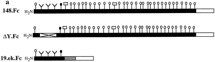
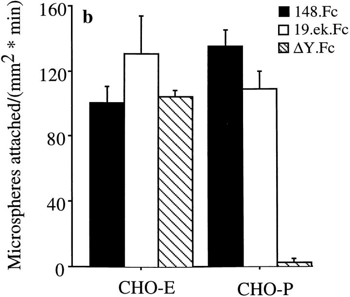
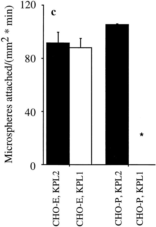
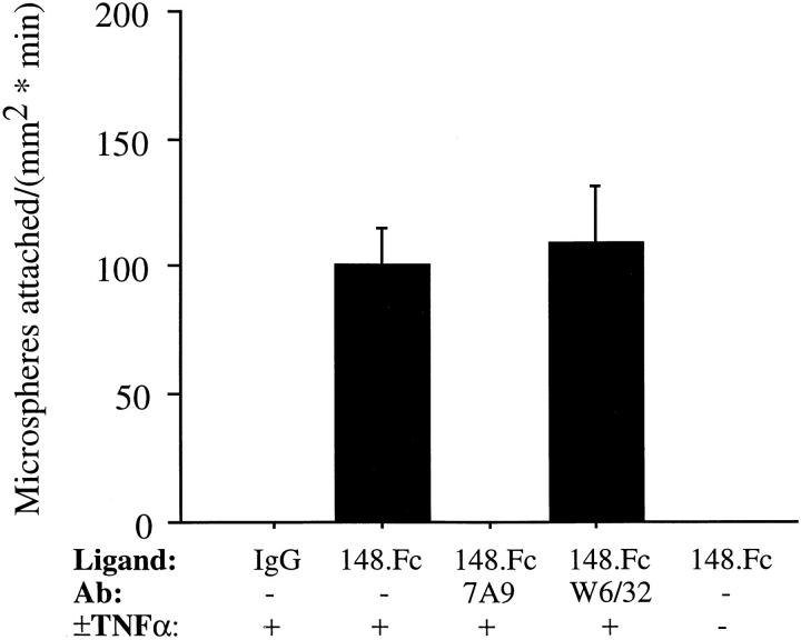
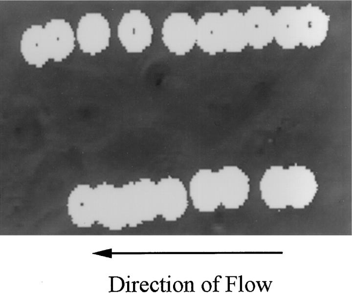
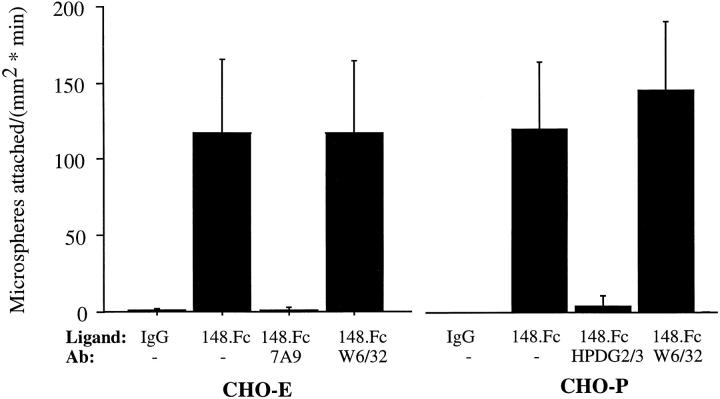
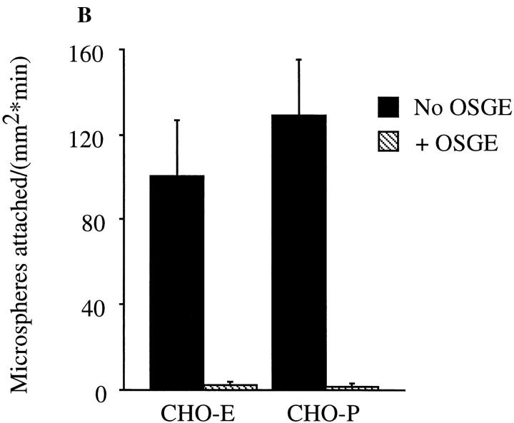
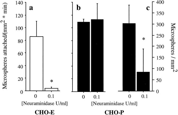
Leukocyte adhesion to vascular endothelium under flow involves an adhesion cascade consisting of multiple receptor pairs that may function in an overlapping fashion. P-selectin glycoprotein ligand-1 (PSGL-1) and L-selectin have been implicated in neutrophil adhesion to P- and E-selectin under flow conditions. To study, in isolation, the interaction of PSGL-1 with P- and E-selectin under flow, we developed an in vitro model in which various recombinant regions of extracellular PSGL-1 were coupled to 10-microm-diameter microspheres. In a parallel plate chamber with well defined flow conditions, live time video microscopy analyses revealed that microspheres coated with PSGL-1 attached and rolled on 4-h tumor necrosis factor-alpha-activated endothelial cell monolayers, which express high levels of E-selectin, and CHO monolayers stably expressing E- or P-selectin. Further studies using CHO-E and -P monolayers demonstrate that the first 19 amino acids of PSGL-1 are sufficient for attachment and rolling on both E- and P-selectin and suggest that a sialyl Lewis x-containing glycan at Threonine-16 is critical for this sequence of amino acids to mediate attachment to E- and P-selectin. The data also demonstrate that a sulfated, anionic polypeptide segment within the amino terminus of PSGL-1 is necessary for PSGL-1-mediated attachment to P- but not to E-selectin. In addition, the results suggest that PSGL-1 has more than one binding site for E-selectin: one site located within the first 19 amino acids of PSGL-1 and one or more sites located between amino acids 19 through 148.


Figures

Figure 6
Figure 6
The first 19 amino acids of PSGL-1 are sufficient for attachment to CHO-E and -P monolayers, and the NH2-terminal anionic polypeptide segment of PSGL-1 is necessary for 148.Fc-mediated attachment to CHO-P, but not CHO-E monolayers. (a) Schematic representation of the PSGL-1.Fc constructs. Closed bars, PSGL-1 segments; open bars, human Fc segments; open bar with X, an internal deletion of amino acids 5–11 within the NH2-terminal anionic polypeptide region; shaded bar, the enterokinase cleavage site; Y, amino-terminal tyrosine; vertical lines with open circles, the approximate number and location of potential O-linked glycosylation sites; vertical lines with closed circles, O-linked site at amino terminal threonine 16; vertical lines with shaded rectangles, potential N-linked glycosylation sites. (Drawing not to scale.) (b) Microspheres coated with the 19.ek.Fc mutant (open bars), containing the first 19 amino acids of PSGL-1, attached to CHO-P and -E monolayers at a rate similar to 148.Fc microspheres (dark bars). Microspheres coated with the ΔY.Fc mutant, which consists of the first 148 amino acids of PSGL-1 with an internal deletion of the residues in positions 5–11, attached to CHO-E monolayers at a rate similar to 148.Fc microspheres but did not attach to CHO-P monolayers (crosshatched bars). (Shear stress = 2 dynes/cm2; n = 2). (c) An mAb to PSGL-1, KPL1, which requires amino acids 5–11 to recognize PSGL-1 (Table I), did not affect attachment of 148.Fc microspheres to CHO-E monolayers but eliminated attachment to CHO-P monolayers. 148.Fc microspheres treated with mAb KPL1 attached to CHO-E monolayers at a rate similar to that observed for 148.Fc microspheres treated with control mAb KPL2 but did not attach to CHO-P monolayers. (Shear stress = 2 dynes/cm2; *P < 0.05; n = 2). (d) mAb KPL1 eliminated attachment of 19.ek.Fc microspheres to both CHO-E and -P monolayers. (Shear stress = 2 dynes/cm2; *P < 0.05; n = 2).
Figure 6
Figure 6
The first 19 amino acids of PSGL-1 are sufficient for attachment to CHO-E and -P monolayers, and the NH2-terminal anionic polypeptide segment of PSGL-1 is necessary for 148.Fc-mediated attachment to CHO-P, but not CHO-E monolayers. (a) Schematic representation of the PSGL-1.Fc constructs. Closed bars, PSGL-1 segments; open bars, human Fc segments; open bar with X, an internal deletion of amino acids 5–11 within the NH2-terminal anionic polypeptide region; shaded bar, the enterokinase cleavage site; Y, amino-terminal tyrosine; vertical lines with open circles, the approximate number and location of potential O-linked glycosylation sites; vertical lines with closed circles, O-linked site at amino terminal threonine 16; vertical lines with shaded rectangles, potential N-linked glycosylation sites. (Drawing not to scale.) (b) Microspheres coated with the 19.ek.Fc mutant (open bars), containing the first 19 amino acids of PSGL-1, attached to CHO-P and -E monolayers at a rate similar to 148.Fc microspheres (dark bars). Microspheres coated with the ΔY.Fc mutant, which consists of the first 148 amino acids of PSGL-1 with an internal deletion of the residues in positions 5–11, attached to CHO-E monolayers at a rate similar to 148.Fc microspheres but did not attach to CHO-P monolayers (crosshatched bars). (Shear stress = 2 dynes/cm2; n = 2). (c) An mAb to PSGL-1, KPL1, which requires amino acids 5–11 to recognize PSGL-1 (Table I), did not affect attachment of 148.Fc microspheres to CHO-E monolayers but eliminated attachment to CHO-P monolayers. 148.Fc microspheres treated with mAb KPL1 attached to CHO-E monolayers at a rate similar to that observed for 148.Fc microspheres treated with control mAb KPL2 but did not attach to CHO-P monolayers. (Shear stress = 2 dynes/cm2; *P < 0.05; n = 2). (d) mAb KPL1 eliminated attachment of 19.ek.Fc microspheres to both CHO-E and -P monolayers. (Shear stress = 2 dynes/cm2; *P < 0.05; n = 2).
Figure 1
Figure 1
A PSGL-1.Fc chimera molecule can be coupled to protein A microspheres. 10-μm microspheres were precoated with protein A. These microspheres were then incubated with various concentrations of the 148.Fc chimera. The amount of 148.Fc chimera bound to the microspheres was detected with an mAb to PSGL-1 (a–e), PSL-275, and an appropriate FITC-labeled secondary antibody. Isotype-matched control mAb to ICAM-1, Hu5/3, did not recognize the 148.Fc chimera (f   ).
Figure 2
Figure 2
148.Fc microspheres attached and rolled on TNF-α–activated HUVEC monolayers under flow. 148.Fc or IgG microspheres were perfused across TNF-α– (4 h, 25 ng/ml) activated or –unactivated HUVEC monolayers. 148.Fc microspheres attached and rolled on TNF-α–activated HUVEC monolayers while IgG microspheres did not attach to the TNF-α–activated HUVEC monolayers. 148.Fc microsphere attachment was blocked by an mAb to E-selectin (7A9) but unaffected by a negative isotypematched control endothelial cell binding mAb to Class I (W6/32). 148.Fc microspheres did not attach to unactivated HUVEC monolayers. (Shear stress = 2 dynes/cm2; n = 3).
Figure 3
Figure 3
148.Fc microspheres rolled on TNF-α–stimulated HUVEC. The image shows two 148.Fc microspheres (white spheres) rolling over a TNF-α–activated HUVEC monolayer (gray background). Images were captured, every 0.6 s, from a videotape of the experiment and layered together to give the composite image shown. Note that the 148.Fc microspheres translated in the direction of the flow with a nonconstant velocity. The average velocity of 10 different 148.Fc microspheres was determined and found to be 14 μm/sec, which is <3% of the hydrodynamic velocity of a noninteracting hard sphere translating 50 nm from the surface (13). The length of the image shown is 100 μm. Shear stress = 2 dynes/cm2.
Figure 4
Figure 4
148.Fc microspheres attached and rolled on CHO-P and -E monolayers. 148.Fc microspheres attached and rolled on CHO-E and -P monolayers while IgG microspheres did not attach to either CHO monolayer. 148.Fc microsphere attachment to CHO-E and -P monolayers was blocked by an mAb to E-selectin (7A9) and an mAb to P-selectin (HPDG2/3), respectively, but was unaffected by an isotype-matched control mAb (W6/32). 148.Fc microspheres did not attach to the parental CHO cell line (data not shown). (Shear stress = 2 dynes/ cm2; n = 3).
Figure 5
Figure 5
OSGE abolishes 148.Fc microsphere attachment to CHO-P or -E monolayers. (A) 148.Fc microspheres were incubated with buffer (a and c) or OSGE (b and d) at 37°C for 30 min. Untreated (a) or OSGE-treated (b) 148.Fc microspheres were incubated with an mAb to PSGL-1 (PSL-275) (open histograms) or an isotype-matched control mAb to ICAM-1 (Hu5/3) (shaded histograms) and subsequently an FITC-labeled secondary antibody. As a control for the specificity of OSGE, the presence of the human Fc region of the 148.Fc chimera was detected on the 148.Fc microspheres. Untreated (c) or OSGE-treated (d) 148.Fc microspheres were incubated with an FITC-labeled polyclonal antibody to human Fc (open histograms) or control, mouse IgG (shaded histograms). Results shown are representative of n = 2–4 separate experiments. (B) Treatment of 148.Fc microspheres with OSGE before use in the in vitro flow assay abolished 148.Fc microsphere attachment to CHO-E and -P monolayers. (Shear stress = 2 dynes/cm2; P < 0.05; n = 3).
Figure 5
Figure 5
OSGE abolishes 148.Fc microsphere attachment to CHO-P or -E monolayers. (A) 148.Fc microspheres were incubated with buffer (a and c) or OSGE (b and d) at 37°C for 30 min. Untreated (a) or OSGE-treated (b) 148.Fc microspheres were incubated with an mAb to PSGL-1 (PSL-275) (open histograms) or an isotype-matched control mAb to ICAM-1 (Hu5/3) (shaded histograms) and subsequently an FITC-labeled secondary antibody. As a control for the specificity of OSGE, the presence of the human Fc region of the 148.Fc chimera was detected on the 148.Fc microspheres. Untreated (c) or OSGE-treated (d) 148.Fc microspheres were incubated with an FITC-labeled polyclonal antibody to human Fc (open histograms) or control, mouse IgG (shaded histograms). Results shown are representative of n = 2–4 separate experiments. (B) Treatment of 148.Fc microspheres with OSGE before use in the in vitro flow assay abolished 148.Fc microsphere attachment to CHO-E and -P monolayers. (Shear stress = 2 dynes/cm2; P < 0.05; n = 3).
Figure 7
Figure 7
Effect of neuraminidase treatment of 19.ek.Fc microsphere adhesive interactions with CHO-E and -P monolayers. Microspheres were coated with 50 μg/ml 19.ek.Fc, treated with neuraminidase, and perfused over CHO-E (open bars) or CHO-P (filled bars) monolayers. (a) The rate of attachment to CHO-E monolayers was significantly diminished, if not eliminated, by treatment with neuraminidase. (b) In contrast, the rate of attachment to CHO-P monolayers was unaffected by neuraminidase treatment. (c) Accumulation of 19.ek.Fc microspheres on CHO-P monolayers was significantly diminished by treatment with neuraminidase. (Shear stress = 2 dynes/cm2; *P < 0.05; n = 3).

References

    1. Abbassi O, Kishimoto TK, McIntire LV, Anderson DC, Smith CW. E-selectin supports neutrophil rolling in vitro under conditions of flow. J Clin Invest. 1993;92:2719–2730. - PMC - PubMed
    1. Alon R, Rossitier H, Wang X, Springer TA, Kupper TS. Distinct cell surface ligands mediate T lymphocyte attachment and rolling on P- and E-selectin under physiological flow. J Cell Biol. 1994;127:1485–1495. - PMC - PubMed
    1. Alon R, Hammer DA, Springer TA. Lifetime of the P-selectin-carbohydrate bond and its response to tensile force in hydrodynamic flow. Nature (Lond) 1995;374:539–542. - PubMed
    1. Arbones ML, Ord DC, Ley K, Radech H, Maynard-Curry C, Otten G, Capon DJ, Tedder TF. Lymphocyte homing and leukocyte rolling and migration are impaired in L-selectin (CD62L) deficient mice. Immunity. 1994;1:247–260. - PubMed
    1. Asa D, Raycroft L, Ma L, Aeed PA, Kaytes PS, Elhammer AP, Geng J. The P-selectin glycoprotein ligand functions as a common human leukocyte ligand for P- and E-selectins. J Biol Chem. 1995;270:11662–11670. - PubMed

Publication types

MeSH terms