. 1997 May 15;17(10):3568-79.
doi: 10.1523/JNEUROSCI.17-10-03568.1997.

Localized and transient elevations of intracellular Ca2+ induce the dedifferentiation of axonal segments into growth cones

Affiliations

Localized and transient elevations of intracellular Ca2+ induce the dedifferentiation of axonal segments into growth cones

N E Ziv et al. J Neurosci. .

Abstract

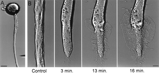
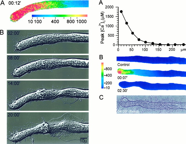
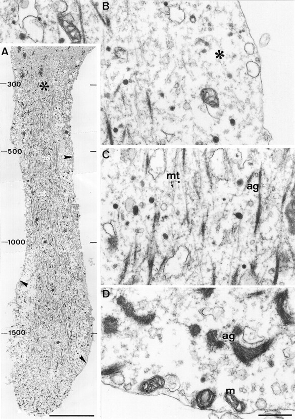
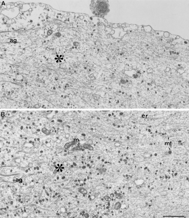
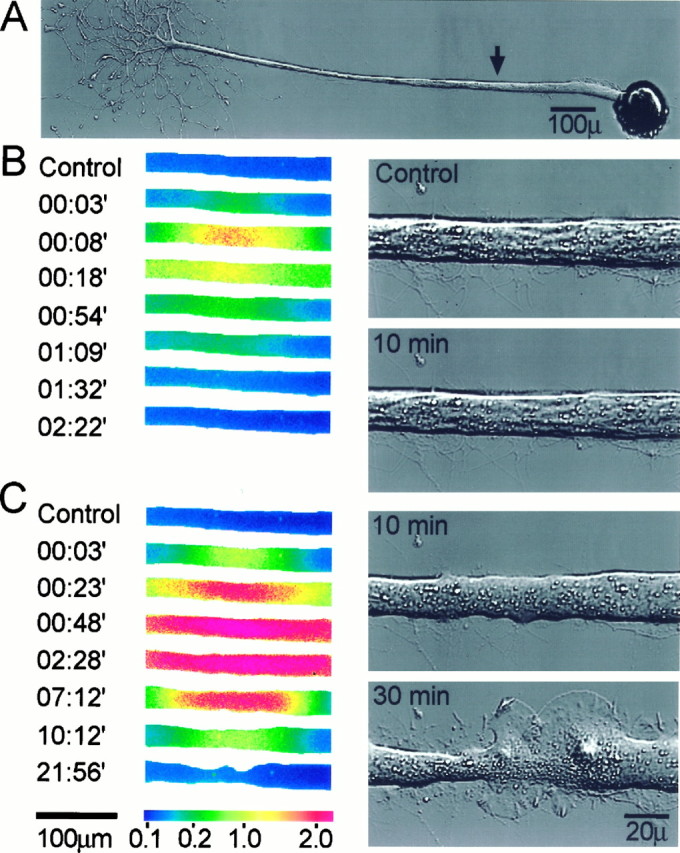
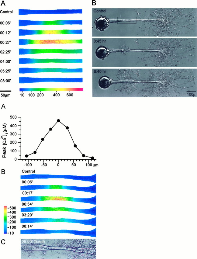
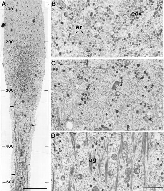
The formation of a growth cone at the tip of a severed axon is a key step in its successful regeneration. This process involves major structural and functional alterations in the formerly differentiated axonal segment. Here we examined the hypothesis that the large, localized, and transient elevation in the free intracellular calcium concentration ([Ca2+]i) that follows axotomy provides a signal sufficient to trigger the dedifferentiation of the axonal segment into a growth cone. Ratiometric fluorescence microscopy and electron microscopy were used to study the relations among spatiotemporal changes in [Ca2+]i, growth cone formation, and ultrastructural alterations in axotomized and intact Aplysia californica neurons in culture. We report that, in neurons primed to grow, a growth cone forms within 10 min of axotomy near the tip of the transected axon. The nascent growth cone extends initially from a region in which peak intracellular Ca2+ concentrations of 300-500 microM are recorded after axotomy. Similar [Ca2+]i transients, produced in intact axons by focal applications of ionomycin, induce the formation of ectopic growth cones and subsequent neuritogenesis. Electron microscopy analysis reveals that the ultrastructural alterations associated with axotomy and ionomycin-induced growth cone formation are practically identical. In both cases, growth cones extend from regions in which sharp transitions are observed between axoplasm with major ultrastructural alterations and axoplasm in which the ultrastructure is unaltered. These findings suggest that transient elevations of [Ca2+]i to 300-500 microM, such as those caused by mechanical injury, may be sufficient to induce the transformation of differentiated axonal segments into growth cones.


Figures

Fig. 1.
Fig. 1.
Axotomy is followed by the rapid formation of a growth cone. A, A low-magnification image of a cultured buccal neuron B1 acquired before axonal transection. The micropipette used for transecting the axon is seen in the bottom right corner. B, After axotomy the morphology of the distal axonal segment was altered, and this was followed by the rapid formation of a growth cone in the form of an extending lamellipodium. Scale bars: A, 50 μm; B, 10 μm.
Fig. 2.
Fig. 2.
(Left) The correlation between peak [Ca2+]i after axotomy and the site of growth cone formation. A mag–fura-2 ratio image of the spatial distribution of [Ca2+]i at the time at which [Ca2+]i was elevated to its maximal levels (A) is compared with DIC images of the same neuron that show the subsequent formation of the new growth cone (B). [Ca2+]i was elevated to ∼300 μm at the site from which the lamellipodium was first observed to extend. [Ca2+]i is given in μm. Time is given in minutes from axotomy.
Fig. 4.
Fig. 4.
The elevation of [Ca2+]ito >300 μm is associated with significant alterations in the axonal cytoarchitecture. A longitudinal section through the distal region of the transected axon shown in Figure 3 reveals large changes in the axonal ultrastructure along a segment of ∼80 μm from the tip of the transected axon, in which [Ca2+]i was elevated to >300 μm. A, A low-magnification view of the transected axon. Note the disruption of microtubules and neurofilaments in the distal region of the axon, the formation of short fragments of electron-dense filamentous material in the core of the axoplasm, and the conspicuous separation of the axolemma from the axoplasmic core (arrowheads). In particular, note the sharp transition (asterisk) between the severely altered axoplasm and the unaltered axoplasm of the proximal region in which [Ca2+]i was elevated to <300 μm. The peak Ca2+ concentrations (in μm) recorded along the axon after axotomy are indicated on the left side of the figure. B, A high-magnification view of the region adjacent to the axolemma reveals a large gap between the axolemma and the cytoskeletal core (asterisk), which is filled with amorphous axoplasm and several large vacuoles. C, A high-magnification view of the severely altered axoplasmic core reveals that the electron-dense filaments are aggregates of amorphous material and short segments of microtubules (ag). D, A high-magnification view of the tip of the cut axon reveals that large electron-dense aggregates are formed near the tip as well as vacuoles of unidentified origin. mt, Microtubules;m, mitochondria. Scale bars: A, 10 μm;BD, 0.5 μm.
Fig. 5.
Fig. 5.
The ultrastructure at the transition zone.A, Axolemma reattachment. A high-magnification view of the axolemma at the transition zone (asterisk) reveals that the axolemma, detached from the axoplasmic core along the distal region of the transected axon (left side), reattaches to the axoplasmic core in this region. B, The abrupt ultrastructural transition takes place over ∼5 μm. As seen in Figures 3 and 4, [Ca2+]i was elevated in this region after axotomy to ∼300 μm. Note the disappearance of the electron-dense aggregates (ag) and the recovery of the linear organization of the axoplasm in the proximal region (right side). mt, Microtubules;m, mitochondria; er, endoplasmic reticulum. Scale bar, 1 μm.
Fig. 6.
Fig. 6.
Transient elevations in [Ca2+]i induce the formation of ectopic growth cones along intact axons. Focal applications of the Ca2+ ionophore ionomycin were used to elevate transiently the [Ca2+]i in intact axons, and the effects of these Ca2+ transients on axonal morphology were recorded. A, The site of ionomycin application (arrow). B, Fura-2 ratio images (left panels) showing the spatiotemporal alterations in the axonal [Ca2+]i induced by a brief ionophore application. [Ca2+]i was elevated transiently to several micromolars. The right panelsshow the axon before (top panel) and 10 min after the application (bottom panel). No significant alterations were caused to the morphology of the axon.C, A second, prolonged ionophore application to the same site elevated [Ca2+]i to levels that exceeded those that could be determined reliably with fura-2 (left panels). The right panels show the axon 10 and 30 min after the second application. Note the general change in the appearance of the axon 10 min after the ionophore application and the subsequent formation of two prominent, overlapping growth cones on both sides of the application site. [Ca2+]i is given in μm. Time is give in minutes from ionomycin application.
Fig. 7.
Fig. 7.
(Top) Axonal dedifferentiation requires transient elevations of [Ca2+]i to 300–600 μm. Mag–fura-2 ratiometric fluorescence microscopy was used to determine the intra-axonal [Ca2+]irequired for inducing the transformation of an intact axonal segment into a growth cone. A, The spatiotemporal alterations in the axonal [Ca2+]i induced by a focal application of ionomycin. The region shown corresponds to therectangle in B, top panel. [Ca2+]i is given in μm.B, The resulting changes in axonal morphology. The transient increase of [Ca2+]i to ∼500 μm induced the formation of a growth cone at the application site, which subsequently developed into a new neuritic tree.
Fig. 9.
Fig. 9.
Local elevation of [Ca2+]i to >300 μm induces ultrastructural alterations similar to those induced by axotomy. A longitudinal section through the axon shown in Figure 8 reveals that the ionophore-induced Ca2+ elevation caused ultrastructural alterations similar to those observed after axotomy (compare with Fig.4). A, A low-magnification view of the proximal region of the application site. In the central region (bottom), where [Ca2+]i was elevated to ∼300 μm or more, major alterations in axonal cytoarchitecture were observed, which included microtubule and neurofilament disruption and the detachment of the axolemma from the axoplasmic core (arrowheads). In the proximal region (top), the axoplasm retained its normal appearance. Many clear and electron-dense vesicles are seen in the transition zone between these two compartments. The peak Ca2+concentrations (in μm) recorded along the axon after the ionophore application are indicated at the left of the figure. B, A high-magnification view of the region immediately proximal to the transition zone shows that it contains a large number of electron-dense vesicles (edv).C, The transition zone. D, A high-magnification view of the short fragments of electron-dense filamentous aggregates (ag) seen in the core of the axoplasm in which [Ca2+]i was elevated to ∼300 μm or more. er, Endoplasmic reticulum. Scale bars: A, 10 μm; D, 1 μm.

References

    1. Adler EM, Augustine GJ, Duffy SN, Charlton MP. Alien intracellular calcium chelators attenuate neurotransmitter release at the squid giant synapse. J Neurosci. 1991;11:1496–1507. - PMC - PubMed
    1. Ashery U, Penner R, Spira ME. Acceleration of membrane recycling by axotomy of cultured Aplysia neurons. Neuron. 1996;16:641–651. - PubMed
    1. Baas PW, Heidemann SR. Microtubule reassembly from nucleating fragments during the regrowth of amputated neurites. J Cell Biol. 1986;103:917–927. - PMC - PubMed
    1. Baas PW, White LA, Heidemann SR. Microtubule polarity reversal accompanies regrowth of amputated neurites. Proc Natl Acad Sci USA. 1987;84:5272–5276. - PMC - PubMed
    1. Bailey CH, Kandel ER. Structural changes accompanying memory storage. Annu Rev Physiol. 1993;55:397–426. - PubMed

Publication types

LinkOut - more resources