. 1997 May 15;17(10):3675-83.
doi: 10.1523/JNEUROSCI.17-10-03675.1997.

Failed cell migration and death of purkinje cells and deep nuclear neurons in the weaver cerebellum

Affiliations

Failed cell migration and death of purkinje cells and deep nuclear neurons in the weaver cerebellum

S M Maricich et al. J Neurosci. .

Abstract

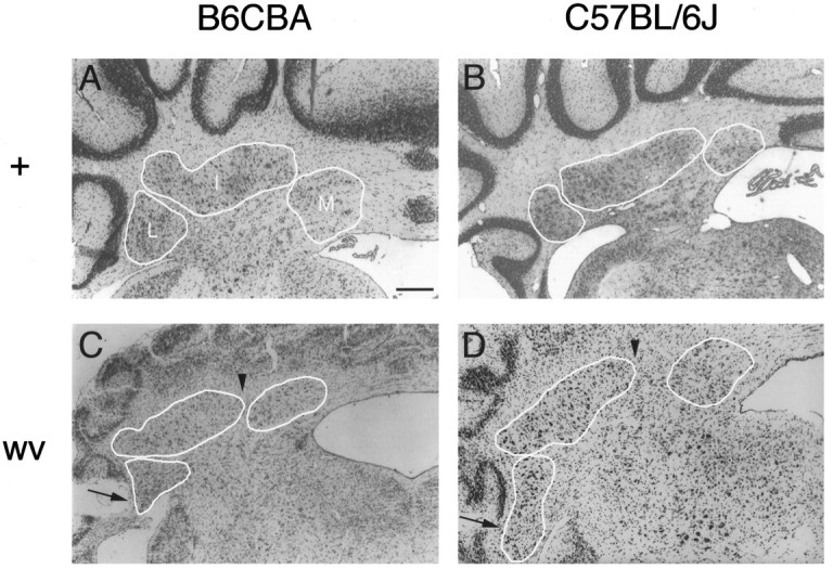
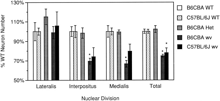
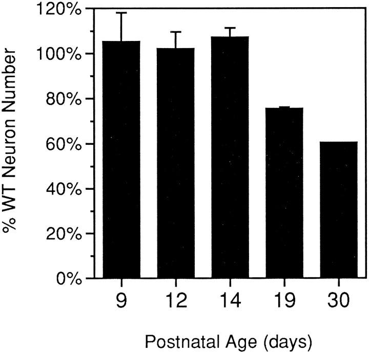
The mouse neurological mutant weaver has an atrophic cerebellar cortex with deficits in both Purkinje and granule cell number. Although granule cells are known to die postnatally shortly after their final cell division, the cause of the Purkinje cell deficit (cell death vs lack of production) is unknown. We report here a quantitative analysis of large cerebellar neurons of the weaver mutant during postnatal development. We explored the hypothesis that the cells of the entire cerebellar anlage were affected by the mutation by including in our study the neurons of the deep cerebellar nuclei (DCN). Our analysis reveals that in homozygous weaver mutants (1) the DCN are displaced laterally, display an abnormal anatomy, and suffer a 20-25% decrease in neuron number; (2) this numerical deficit is located in medial regions, similar to the localization of cortical deficits in both Purkinje and granule cells; (3) pyknotic figures are present in the juvenile DCN and in the Purkinje cell layer; and (4) the majority of cell death in these populations occurs not in medial regions where the numerical deficits are observed, but rather laterally where adult cell number is nearly normal. These results lead us to propose that the complete weaver phenotype includes a failure of the cell movements that lead to the fusion of the bilateral cerebellar anlage, and that this failure to migrate properly leaves some of the Purkinje cells and DCN neurons in a position where they are unable to make appropriate connections, leading to their death. In addition to implications for normal development, these observations suggest that weaver effects on the cerebellum can be unified into one consolidated model in which failure of cell movement affects all major cerebellar neurons.


Figures

Fig. 1.
Fig. 1.
The weaver mutation affects the basic morphology of the DCN in C57BL/6J and B6CBA mouse strains. All photographs are at 50× magnification from approximately the same rostrocaudal level of adult animals; individual nuclei are outlined and indicated on each photo. A, B6CBA +/wvheterozygote. Wild-type B6CBA DCN (not shown) appear virtually identical to the DCN of the heterozygote. B, Wild-type C57BL/6J DCN. Note that nucleus interpositus is slightly more elongated compared with B6CBA DCN. C, B6CBA wv/wvmutant. Arrows point to nucleus lateralis neurons in the white matter of the paraflocculus, a condition that is absent in wild-type and heterozygous mice. Arrowheads illustrate the more lateral location of the medial boundary of nucleus interpositus. D, C57BL/6J wv/wv mutant. Although larger in absolute size than the B6CBA homozygous mutant, the basic DCN anatomy is unaltered. Arrows andarrowheads same as C. Scale bar, 200 μm.
Fig. 2.
Fig. 2.
The weaver mutation selectively affects medial portions of the DCN, causing a decrease in total neuron number. Neuron counts are graphed as a percentage of wild type on the same strain background; absolute numbers for these animals are presented in Table 1. Asterisks denote difference from wild-type values that is statistically significant (ANOVA, Newman–Keuls; p < 0.05). Error bars represent SEM.
Fig. 3.
Fig. 3.
Comparison of DCN neuron distribution in C57BL/6J wild type and weaver. Cell counts were taken every 80 μm in sagittally sectioned brains and then graphed versus percentage mediolateral distance from the most medial aspect of the DCN. One side of one wild-type and both sides of one weaver brain are shown. Note the selective decrease in medial counts, particularly those in medial nucleus interpositus, in the weaver DCN.Bars at the top of the figure indicate approximate boundaries of each nucleus as determined by measurement of nuclei in several coronal sections.
Fig. 4.
Fig. 4.
Juvenile weaver mutants contain dying cells in the DCN and Purkinje cell layer. Photographs are of one lateral section of a C57BL/6J P14 weaver cerebellum sectioned sagittally. Rostral is to the right in all photographs. A, Lateral cerebellum at 50×. White boxes correspond to views in B andC. B, DCN at 400×.Arrowhead and asterisk indicate a dying cell seen at 1000× in inset. C, Cerebellar cortex at 400×. Two putative pyknotic Purkinje cells are indicated by arrowheads; inset is 1000× photo of cell marked by asterisk. Pyknotic Purkinje cells were not observed in wild-type cortex at any age (data not shown). Scale bars: A, 200 μm; B, C, 50 μm; insets, 5 μm.
Fig. 5.
Fig. 5.
Number of pyknoses in the DCN of juvenileweaver mutants is higher than that found in wild-type mice. The number of dying cells was determined for C57BL/6J animals every 80 μm in sagittally sectioned brains (one side of one brain of each age). The DCN was then divided into four regions of equal size by percentage of total mediolateral distance, and the counts were summed for each of these regions. A, Wild-type DCN pyknoses. Dying cells are relatively rare and evenly distributed across the mediolateral extent of the DCN. B, weaverDCN pyknoses. Pyknotic figures are seen in greater numbers in lateral portions of the DCN. The largest number of dying cells is seen atP19.
Fig. 6.
Fig. 6.
Medium-to-large neuron (MLN) number decreases laterally in weaver mutants between P9 and P19. The number of MLNs was counted every 160 μm in sagittal sections of C57BL/6J cerebella and graphed versus distance from the midline. Counts from one side of three wild-type animals (solid lines) and four weaver cerebella (dotted lines) are shown. At P9, weaver mice have many more MLNs laterally than do wild-type mice. These numbers gradually decline toward wild-type levels by P19. No change takes place in medial MLN numbers.
Fig. 7.
Fig. 7.
Total MLN number decreases from wild-type levels as weaver animals age. Mean MLN counts (average of both sides of the same brain) are graphed as a percentage of wild-type counts at corresponding ages; P9–P19 brains are the same as those shown in Figure 6. Error bars represent SEM. Data for the P30weaver were obtained from Herrup and Trenkner (1987).

References

    1. Altman J, Anderson WJ. Experimental reorganization of the cerebellar cortex. I. Morphological effects of elimination of all microneurons with prolonged x-irradiation started at birth. J Comp Neurol. 1972;146:355–406. - PubMed
    1. Altman J, Bayer S. Embryonic development of the rat cerebellum. I. Delineation of the cerebellar primordium and early cell movements. J Comp Neurol. 1985a;231:1–26. - PubMed
    1. Altman J, Bayer S. Embryonic development of the rat cerebellum. II. Translocation and regional distribution of the deep neurons. J Comp Neurol. 1985b;231:27–41. - PubMed
    1. Altman J, Bayer S. Embryonic development of the rat cerebellum. III. Regional differences in the time of origin, migration and settling of Purkinje cells. J Comp Neurol. 1985c;231:42–65. - PubMed
    1. Armstrong DM, Schild RF. An investigation of the cerebellar cortico-nuclear projections in the rat using an autoradiographic tracing method. I. Projection from the vermis. Brain Res. 1978a;141:1–19. - PubMed

Publication types

MeSH terms

Substances

LinkOut - more resources