. 1997 May 15;17(10):3727-38.
doi: 10.1523/JNEUROSCI.17-10-03727.1997.

Dentate granule cell neurogenesis is increased by seizures and contributes to aberrant network reorganization in the adult rat hippocampus

Affiliations

Dentate granule cell neurogenesis is increased by seizures and contributes to aberrant network reorganization in the adult rat hippocampus

J M Parent et al. J Neurosci. .

Abstract

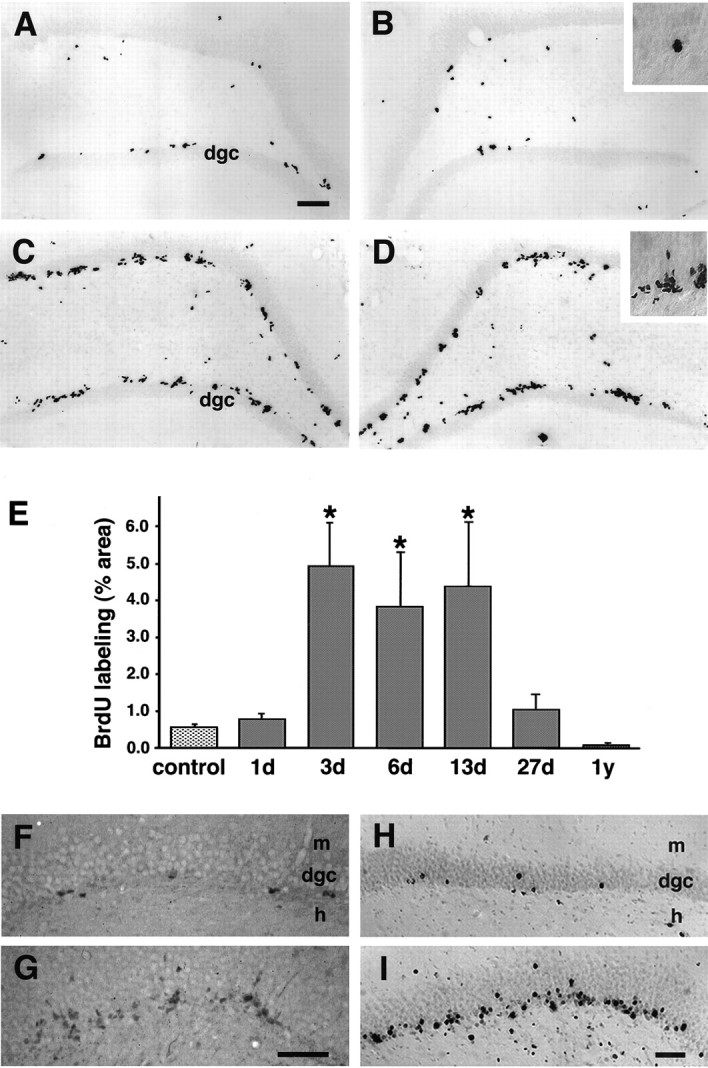
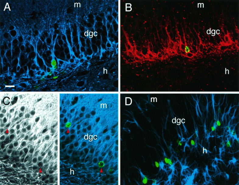
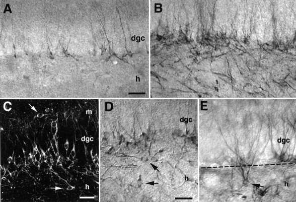
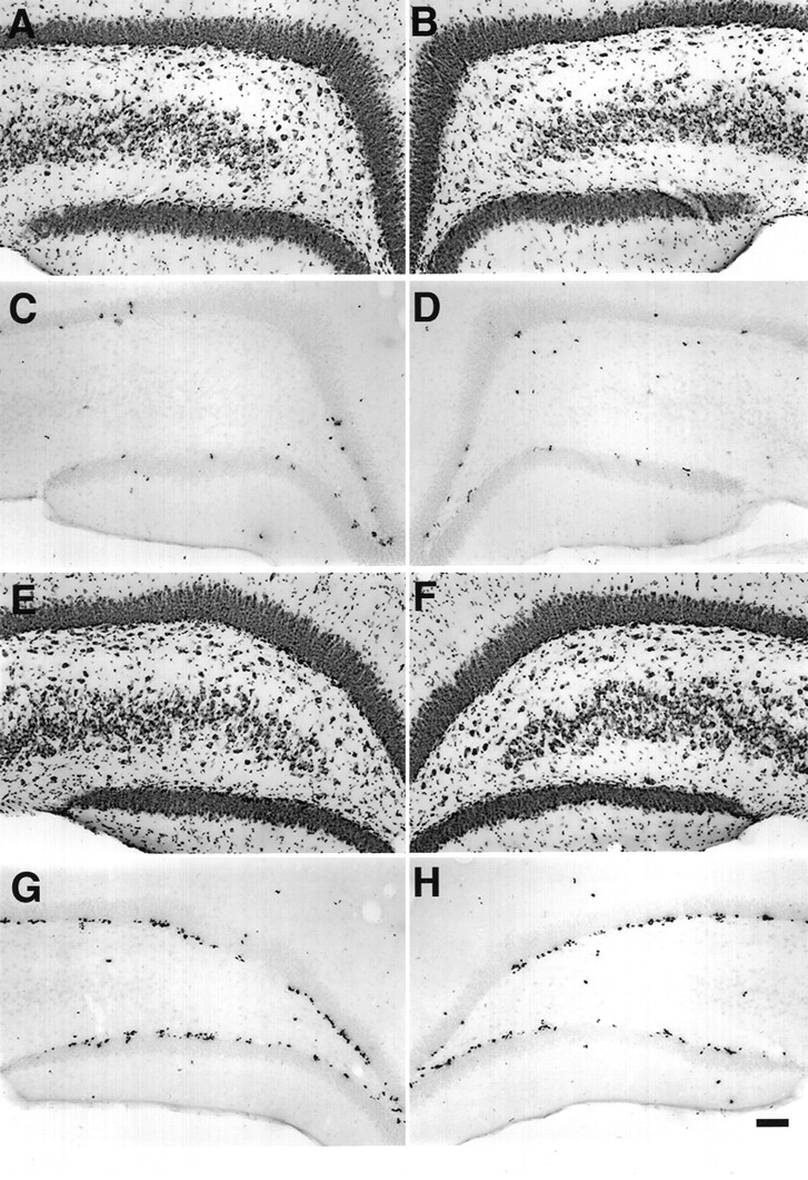
The dentate granule cell layer of the rodent hippocampal formation has the distinctive property of ongoing neurogenesis that continues throughout adult life. In both human temporal lobe epilepsy and rodent models of limbic epilepsy, this same neuronal population undergoes extensive remodeling, including reorganization of mossy fibers, dispersion of the granule cell layer, and the appearance of granule cells in ectopic locations within the dentate gyrus. The mechanistic basis of these abnormalities, as well as their potential relationship to dentate granule cell neurogenesis, is unknown. We used a systemic chemoconvulsant model of temporal lobe epilepsy and bromodeoxyuridine (BrdU) labeling to investigate the effects of prolonged seizures on dentate granule cell neurogenesis in adult rats, and to examine the contribution of newly differentiated dentate granule cells to the network changes seen in this model. Pilocarpine-induced status epilepticus caused a dramatic and prolonged increase in cell proliferation in the dentate subgranular proliferative zone (SGZ), an area known to contain neuronal precursor cells. Colocalization of BrdU-immunolabeled cells with the neuron-specific markers turned on after division, 64 kDa, class III beta-tubulin, or microtubule-associated protein-2 showed that the vast majority of these mitotically active cells differentiated into neurons in the granule cell layer. Newly generated dentate granule cells also appeared in ectopic locations in the hilus and inner molecular layer of the dentate gyrus. Furthermore, developing granule cells projected axons aberrantly to both the CA3 pyramidal cell region and the dentate inner molecular layer. Induction of hippocampal seizure activity by perforant path stimulation resulted in an increase in SGZ mitotic activity similar to that seen with pilocarpine administration. These observations indicate that prolonged seizure discharges stimulate dentate granule cell neurogenesis, and that hippocampal network plasticity associated with epileptogenesis may arise from aberrant connections formed by newly born dentate granule cells.


Figures

Fig. 1.
Fig. 1.
Upregulation of cell proliferation in the adult dentate gyrus after status epilepticus. A, B, Baseline mitotic activity in the dentate gyrus of a saline-treated control rat identified by BrdU labeling and immunohistochemistry. C, D, Increased dentate SGZ BrdU incorporation 13 d after pilocarpine-induced status epilepticus. Note the clustering of BrdU-IR nuclei in the SGZ at the border of the hilus and granule cell layer (insets in B and D). In both animals, BrdU immunohistochemistry was performed 24 hr after the animal received four intraperitoneal injections of 50 mg/kg BrdU over 6 hr. E, Proliferative activity in the dentate SGZ and granule cell layer was significantly increased at 3 d, remained elevated at 6 and 13 d, and returned to baseline levels by 27 d after pilocarpine treatment. Proliferative activity is represented as a percentage of the area labeled by BrdU-immunostaining in the SGZ and dentate granule cell layer (mean ± SEM), as determined by quantitative densitometric analysis.Asterisks denote statistically significant differences from controls (p < 0.05; ANOVA with Fisher’s PLSD post hoc test). F, G, PCNA immunostaining in the dentate gyrus of adult rats 4 d after saline (F) or pilocarpine treatment (G) demonstrated an increase in dentate SGZ mitotic activity after pilocarpine-induced status epilepticus similar to that seen with BrdU labeling. H, I, Delayed BrdU immunostaining in the dentate gyrus of control (H) and pilocarpine-treated (I) adult rats revealed a separation of labeled nuclei that were previously clustered (seeB, D), and increased numbers of labeled nuclei that appear to have migrated further into the granule cell layer, especially in the pilocarpine-treated animal. BrdU was administered on day 7 after saline injection or pilocarpine-induced status epilepticus, and immunohistochemistry was performed 4 weeks later. Scale bars:A–D, 100 μm; F–I, 50 μm.dgc, Dentate granule cell layer; m, molecular layer; h, hilus.
Fig. 2.
Fig. 2.
Neuronal phenotype of BrdU-labeled cells in the dentate granule cell layer after pilocarpine-induced status epilepticus. Animals received BrdU on day 7 after pilocarpine treatment and were killed 7, 14, or 28 d later. A, Nuclear BrdU immunoreactivity (green) colocalized with immunostaining using TuJ1 (blue), a monoclonal antibody against the neuron-specific marker class III β-tubulin, in animals killed 7 d after BrdU injection. Neuron-specific β-tubulin is expressed in early postmitotic and differentiated neurons, and in some mitotically active neuronal precursors. B, Nuclear BrdU-IR (green) was seen within cells immunostained for TOAD-64 (red) 14 d after BrdU injection. TOAD-64 is a membrane-associated marker expressed in the cell bodies and processes of newly born, but not adult, neurons. Note that most of the TOAD-64-IR cells are BrdU-negative because of the abbreviated availability of BrdU for incorporation into S-phase cells after a 6 hr injection period, as compared with the more prolonged accumulation of newly postmitotic TOAD-IR neurons. C,Left panel, Immunofluorescence using antibodies to MAP2 labeled cell bodies and dendrites of dentate granule neurons in an adult rat 28 d after BrdU administration (and 35 days after pilocarpine). Right panel, In the same section, BrdU-IR (green) colocalized with immunostaining for MAP2 (blue) (red arrowheads indicate colocalization). Note that double-labeled cells in A–Cpossess characteristic dentate granule cell morphology (medium-sized nuclei with round or oval-shaped cell bodies, and dendrites extending into the molecular layer). D, In contrast to colocalization of BrdU with neuronal markers, BrdU-IR (green) rarely colocalized with the astrocytic markers vimentin (blue) or GFAP (not shown).A–C are 1 μm optical sections obtained by confocal microscopy to resolve antibody localization within individual cells.D is a composite of 19 stacked optical sections. Scale bar (shown in A): 25 μm. dgc, Dentate granule cell layer; h, hilus; m, molecular layer.
Fig. 3.
Fig. 3.
Status epilepticus alters the location of newly born granule cell bodies and their processes. Immunocytochemistry with antibodies to the neuron-specific, early postmitotic marker TOAD-64 revealed newly differentiating neurons in the granule cell layer (dgc) of adult rats 28 d after saline (A) or pilocarpine (B) administration. The number of TOAD-64-immunostained cells increased after status epilepticus, and many TOAD-64-immunolabeled processes exhibited a disorganized pattern that was not seen in controls.C–E, In pilocarpine-treated but not control rats, many immunolabeled cell bodies with the size and shape of granule cells were found in the inner molecular layer (arrow inC) or in the hilus (arrows inC–E). Note in E the presence of a TOAD-64-IR cell in the hilus with a soma and dendritic arbor characteristic of a dentate granule cell, yet ectopically located ∼50 μm from the dgc. The dotted linedemarcates the DGC/hilar border. Scale bars: A, B, 100 μm; C–E, 40 μm. dgc, Dentate granule cell layer; h, hilus; m, molecular layer.
Fig. 4.
Fig. 4.
Status epilepticus leads to a disruption of normal patterns of newly born granule cell axon outgrowth. A, C, Timm staining of sections from the mid-portion of the hippocampus. Pilocarpine-treated rats demonstrated dense, aberrant reorganization of granule cell mossy fiber terminals into the stratum oriens of the CA3 pyramidal cell region in pilocarpine-treated rats (asterisk in C). This was not typically seen in controls (A), except for occasional mild-to-moderate staining in anterior hippocampal regions. TOAD-64 immunolabeling of similar hippocampal regions in the same control (B) and pilocarpine-treated (D) animals confirmed that the sprouting involved the outgrowth of TOAD-64-immunostained mossy fibers derived from newly postmitotic dentate granule cells (asterisk in D).E, Evidence for the presence of aberrant axons from newly born granule cells in the inner molecular layer of pilocarpine-treated animals. Within the molecular layer, TOAD-IR fibers were seen oriented perpendicular to the normal dendritic pattern of staining (arrowheads). The coexistence of immunoreactive dendrites prohibited the identification of these perpendicular processes as axons. F, G, Double-label immunohistochemistry using antibodies to BrdU and NF-M provided direct evidence for newborn cells sending aberrant axons into the molecular layer. Yellow arrowheads denote BrdU-labeled nuclei of the cells of origin; black arrowheads delineate the trajectory of the NF-M-stained axons. Note the presence of an axonal branch oriented toward the hilus (arrow, G). Scale bar:A–D, 100 μm; E, 50 μm; F, G, 25 μm. dgc, Dentate granule cell layer;h, hilus; m, molecular layer.
Fig. 5.
Fig. 5.
A, B, Nissl-stained dentate gyrus of a sham-stimulated animal showing normal structure on the side contralateral (A) and ipsilateral (B) to electrode placement. C, D, BrdU labeling in the same sham control animal. BrdU was injected 6 d after the end of 6 hr sham stimulation, and the animal was killed 1 d after BrdU administration. E, F, Nissl-stained dentate gyrus after 6 hr perforant path stimulation. Note the relatively normal histological structure after this duration of stimulation, which produces little or no damage. G, H, BrdU labeling of sections from the same animal showing that 6 hr of stimulation increased BrdU labeling in the SGZ bilaterally, similar to the pattern of BrdU-IR seen after pilocarpine treatment (Fig. 1C,D). The mean ± SEM of % area BrdU labeled in the SGZ and dentate granule cell layer was 0.69 ± 0.19 for controls (n = 6) and 1.81 ± 0.53 for stimulated animals (n = 6). Scale bar, 100 μm.

References

    1. Altar CA, Baudry M. Systemic injection of kainic acid: gliosis in olfactory and limbic brain regions quantified with [3H]PK 11195 binding autoradiography. Exp Neurol. 1990;109:333–341. - PubMed
    1. Altman J, Das GD. Autoradiographic and histological evidence of postnatal hippocampal neurogenesis in rats. J Comp Neurol. 1965;124:319–335. - PubMed
    1. Altman J, Das GD. Postnatal neurogenesis in the guinea pig. Nature. 1967;214:1098–1101. - PubMed
    1. Babb TL, Kupfer WR, Pretorious JK, Crandall PH, Levesque MF. Synaptic reorganization by mossy fibers in human epileptic fascia dentata. Neuroscience. 1991;42:351–363. - PubMed
    1. Bayer SA, Yackel JW. Neurons in the rat dentate gyrus granular layer substantially increase during juvenile and adult life. Science. 1982;216:890–892. - PubMed

Publication types

LinkOut - more resources