. 1997 May 15;17(10):3804-14.
doi: 10.1523/JNEUROSCI.17-10-03804.1997.

Dynamic control of location-specific information in tactile cutaneous reflexes from the foot during human walking

Affiliations

Dynamic control of location-specific information in tactile cutaneous reflexes from the foot during human walking

B M Van Wezel et al. J Neurosci. .

Abstract

The purpose of the present study was to determine whether tactile cutaneous reflexes from the skin of the foot contain location-specific information during human walking. Muscular responses to non-nociceptive electrical stimulation of the sural, posterior tibial, and superficial peroneal nerves, each supplying a different skin area of the foot, were studied in both legs during walking on a treadmill. For all three nerves the major responses in all muscles were observed at a similar latency of approximately 80-85 msec. In the ipsilateral leg these reflex responses and their phase-dependent modulation were highly nerve-specific. During most of the stance phase, for example, the peroneal and tibial nerves generally evoked small responses in the biceps femoris muscle. In contrast, during late swing large facilitations generally occurred for the peroneal nerve, whereas suppressions were observed for the tibial nerve. In the contralateral leg the reflex responses for the three nerves were less distinct, although some nerve specificity was observed for individual subjects. It is concluded that non-nociceptive stimulation of the sural, posterior tibial, and superficial peroneal nerves each evokes distinct reflex responses, indicating the presence of location-specific information from the skin of the foot in cutaneous reflexes during human walking. It will be argued that differentially controlled reflex pathways can account for the differences in the phase-dependent reflex modulation patterns of the three nerves, which points to the dynamic control of this information during the course of a step cycle.


Figures

Fig. 1.
Fig. 1.
Ipsilateral biceps femoris (iBF) of subject 1.A, iBF background locomotor activity in relation to the stance (signal up) and swing phases of both legs. Stick diagrams are shown on top. One single step cycle is subdivided into 16 equal intervals, the beginning of the first interval coinciding with ipsilateral footfall (see Materials and Methods). B, For the iBF a typical set of 48 average (n = 10 trials) subtraction traces is shown. This set is subdivided into three subsets that refer to the three different nerves that were stimulated (sural, posterior tibial, and superficialperoneal nerves). The solid linesindicate the time of stimulation, whereas the dotted lines refer to a delay of 100 msec with respect to the onset of stimulation. The stippled lines indicate the time windows that are set around the responses (see Materials and Methods). The time windows were set, using the responses of only those phases in which the maximum exceeded 1 SD of the corresponding background activity. Because the maximum is the value of one single data point, the subtracted data first were smoothed (with a second-order low-pass digital Butterworth filter having a cut-off frequency of 50 Hz) so that a reliable determination of the maximum was possible. This maximum was sought within 50 and 130 msec after the stimulus to assure that the responses with a latency of ∼80 msec were certainly within that time scale. It then was checked whether the value of this maximum exceeded 1 SD of the unsmoothed background activity. When this was the case, the subtracted data of that phase were selected for determination of the time window. For this subject the time windows for iBF were 80–110, 80–108, and 82–106 for the sural, tibial, andperoneal nerves, respectively. The numberon the left side of each subtraction trace indicates in which of the 16 step cycle intervals the response with a latency of ∼80 msec occurs. In all panels: time calibration, 100 msec; EMG calibration, 1 mV. Note that for visual purposes the size of the EMG calibration bars differs for the three nerves.
Fig. 2.
Fig. 2.
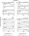
Ipsilateral muscles of subject 1. For the three nerves the subtracted responses (±SE, n = 10 trials) are plotted for all four ipsilateral muscles as a function of the phase in the step cycle. The statistical significance of the reflexes (Wilcoxon signed rank test, p < 0.05) is indicated by the black dots. Phases 1 and9 correspond to the onset of ipsilateral and contralateral stance, respectively. On the top of these figures the background activity of the corresponding muscle is shown. The data are normalized with respect to the maximum background locomotor activity of each muscle (see Materials and Methods). The ipsilateral (i) and contralateral (c) stance phases are shown at the bottom. Note the different vertical scales for the different muscles.
Fig. 3.
Fig. 3.
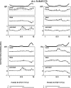
Population average of the ipsilateral muscles (n = 10 subjects). The same format is used as in Figure 2.
Fig. 4.
Fig. 4.
Subtracted ankle angles during the swing phase. On the basis of the dominant iTA modulation pattern (see Fig. 3), kinesiological data were averaged for the sural(n = 9 subjects), tibial(n = 9), and peroneal(n = 7) nerves. Periods are averages of phases 10–11 (early swing), 12–14 (mid-swing), and 15–16 (late swing).Thick solid lines indicate time of stimulation. Thedashed lines are set at 100 msec with respect to each other. Ankle calibration in degrees: PF, plantar flexion; DF, dorsiflexion.
Fig. 5.
Fig. 5.
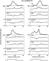
Population average of the contralateral muscles (n = 10 subjects). The same format is used as in Figure 2.
Fig. 6.
Fig. 6.
Contralateral biceps femoris (cBF) of subject 2. The same format is used as in Figure 2.

Similar articles

Cited by

References

    1. Abraham LD, Marks WB, Loeb GE. The distal hindlimb musculature of the cat. Cutaneous reflexes during locomotion. Exp Brain Res. 1985;58:594–603. - PubMed
    1. Aniss AM, Gandevia SC, Burke D. Reflex changes in muscle spindle discharge during a voluntary contraction. J Neurophysiol. 1988;59:908–921. - PubMed
    1. Aniss AM, Gandevia SC, Burke D. Reflex responses in active muscles elicited by stimulation of low-threshold afferents from the human foot. J Neurophysiol. 1992;67:1375–1384. - PubMed
    1. Baker SN, Lemon RN. Non-linear summation of responses in averages of rectified EMG. J Neurosci Methods. 1995;59:175–181. - PubMed
    1. Buford JA, Smith JL. Adaptive control for backward quadrupedal walking. III. Stumbling corrective reactions and cutaneous reflex sensitivity. J Neurophysiol. 1993;70:1102–1114. - PubMed

Publication types