. 1997 May 15;17(10):3894-906.
doi: 10.1523/JNEUROSCI.17-10-03894.1997.

Molecular and physiological diversity of cortical nonpyramidal cells

Affiliations

Molecular and physiological diversity of cortical nonpyramidal cells

B Cauli et al. J Neurosci. .

Abstract

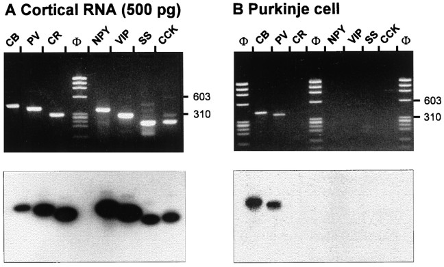
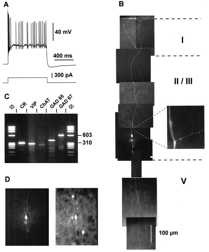
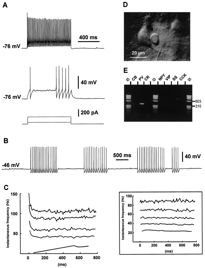
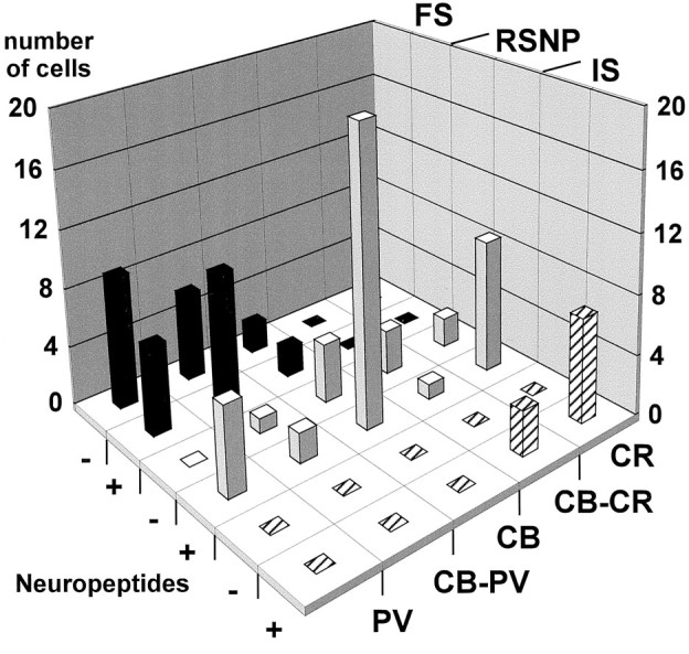
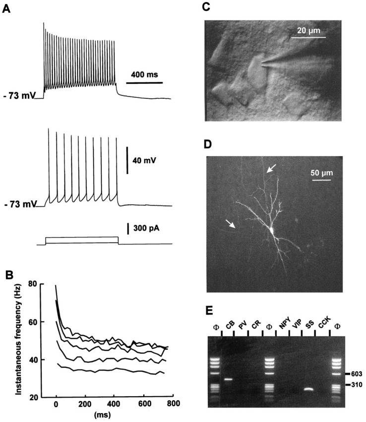
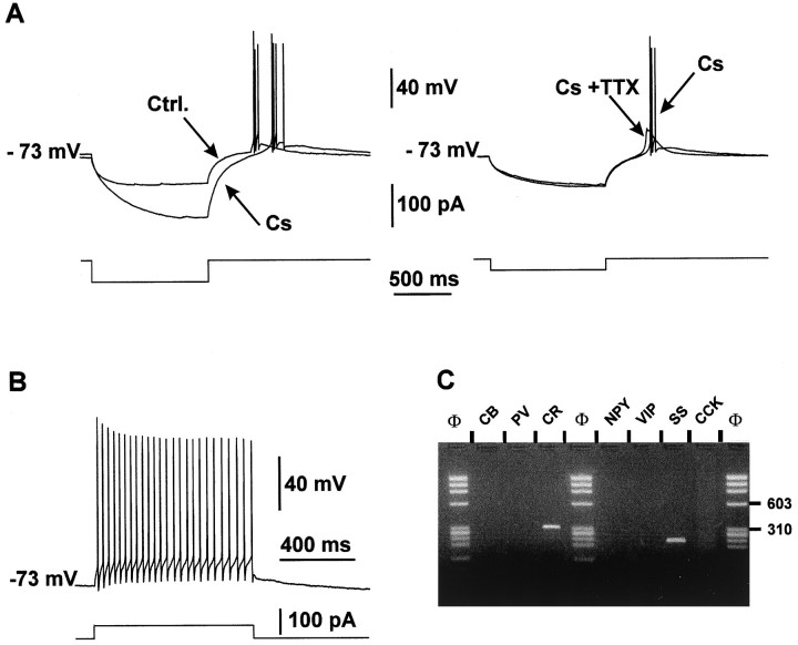
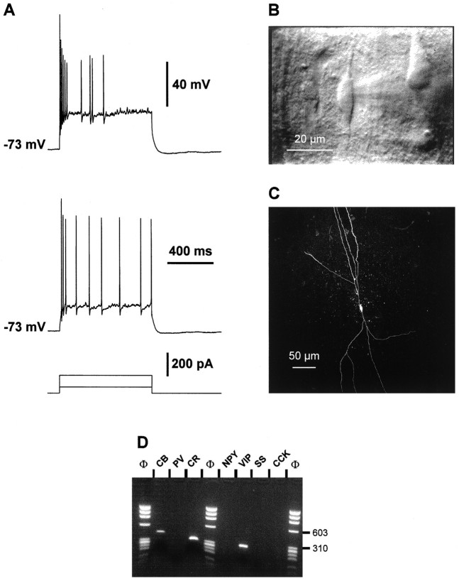
The physiological and molecular features of nonpyramidal cells were investigated in acute slices of sensory-motor cortex using whole-cell recordings combined with single-cell RT-PCR to detect simultaneously the mRNAs of three calcium binding proteins (calbindin D28k, parvalbumin, and calretinin) and four neuropeptides (neuropeptide Y, vasoactive intestinal polypeptide, somatostatin, and cholecystokinin). In the 97 neurons analyzed, all expressed mRNAs of at least one calcium binding protein, and the majority (n = 73) contained mRNAs of at least one neuropeptide. Three groups of nonpyramidal cells were defined according to their firing pattern. (1) Fast spiking cells (n = 34) displayed tonic discharges of fast action potentials with no accommodation. They expressed parvalbumin (n = 30) and/or calbindin (n = 19) mRNAs, and half of them also contained transcripts of at least one of the four neuropeptides. (2) Regular spiking nonpyramidal cells (n = 48) displayed a firing behavior characterized by a marked accommodation and presented a large diversity of expression patterns of the seven biochemical markers. (3) Finally, a small population of vertically oriented bipolar cells, termed irregular spiking cells (n = 15), fired bursts of action potentials at an irregular frequency. They consistently co-expressed calretinin and vasoactive intestinal polypeptide. Additional investigations of these cells showed that they also co-expressed glutamic acid decarboxylase and choline acetyl transferase. Our results indicate that neocortical nonpyramidal neurons display a large diversity in their firing properties and biochemical patterns of co-expression and that both characteristics could be correlated to define discrete subpopulations.


Figures

Fig. 1.
Fig. 1.
Sensitivity and specificity of the RT-mPCR.A, Neocortical RNA (500 pg) was subjected to the RT-mPCR protocol (see Materials and Methods). The seven PCR products were resolved in separate lanes by agarose gel electrophoresis in parallel with Φx174 digested by HaeIII (Φ) as molecular weight marker and stained with ethidium bromide (top panel). The amplified fragments had the sizes (in bp) predicted by the mRNA sequences: 432 (CB), 388 (PV), 309 (CR), 359 (NPY), 287 (VIP), 209 (SS), and 216 (CCK). Southern blot analysis of this agarose gel performed with specific radiolabeled oligoprobes is shown in the bottom panel.B, RT-mPCR applied on a single cerebellar Purkinje cell after electrophysiological recording. Agarose gel electrophoresis (top panel) showed PCR-generated fragments corresponding to CB and PV, as confirmed by Southern blot analysis using specific oligoprobes (bottom panel).
Fig. 6.
Fig. 6.
Physiological, morphological, and molecular analyses of a layer II–III IS cell. A, Current-clamp recording obtained in response to a depolarizing current pulse (300 pA). Note the initial burst followed by irregularly discharged action potentials. B, Reconstruction of the cell labeled with biocytin injection revealed a typical bipolar morphology with two narrow vertical dendritic arborizations. The ascending dendrite (top) extended up to layer I where it branched before reaching the cortical surface. The descending dendrites extended to layer V. Inset, Higher magnification of a portion of the proximal ascending dendrite showing the beginning of the axon running horizontally. Note the ascending and descending colaterals displaying varicosities. C, Agarose gel analysis of products of the RT-mPCR designed for the detection of CR, VIP, ChAT, GAD65, and GAD67 mRNAs. The five amplified fragments had the sizes (in bp) predicted by the mRNA sequences of 309 (CR), 287 (VIP), 262 (ChAT), 391 (GAD65), and 600 (GAD67).D, Comparison of the biocytin labeling of the recorded neuron (left panel) with the immunostaining of the slice with an antibody against CR (right panel). Note the immunoreactivity of the biocytin-labeled cell.
Fig. 2.
Fig. 2.
Electrophysiological and biochemical characterization of a layer V FS cell. A, Current-clamp recording during injection of depolarizing current pulses. Membrane potential was adjusted to −76 mV by continuous current injection, as indicated on the left of each recording. In response to a near-threshold current pulse (50 pA; bottom trace), this FS cell emitted a single fast action potential with a large AHP followed by a silent period and a late discharge of action potentials. Note the membrane potential oscillations during the silent period. Application of a larger depolarizing current (200 pA; top trace) induced a continuous discharge at high frequency.B, Continuous near-threshold depolarizing current (60 pA) evoked clusters of nonaccommodating discharges of fast action potentials. Note the membrane potential oscillations between these clusters. C, Left panel, Analysis of the instantaneous firing frequency during the action potentials discharges evoked in the same FS cell by current pulses of increasing intensities (50–250 pA; bottom to top traces). Note that after a fast early accommodation, discharges rapidly reached a steady-state frequency. Inset, Analysis of the instantaneous firing frequency during the action potentials discharges evoked in another FS cell by current pulses of increasing intensities (50, 100, 150, 200, and 350 pA; bottom to top traces). Note the lack of early accommodation of the discharge frequency at each stimulation intensity. D, IR videomicroscopy picture of the FS cell. The FS cell is located at the center and has a small round soma (diameter ∼10 μm). Note the large layer V pyramidal cell immediately on the left. Pial surface isupward. E, Agarose gel analysis of the RT-mPCR products of the same FS cell. The only PCR-generated fragment was that of PV.
Fig. 7.
Fig. 7.
Expression patterns in different subtypes of neocortical nonpyramidal cells. This histogram shows the distribution of the cells from each physiological subtype according to their CaBP expression and the occurrence of neuropeptides. +, Expression of at least one neuropeptide; −, none of the four neuropeptides expressed. Most of the fast spiking cells (FS, black bars) expressed PV, except four cells that expressed only CB. None of them expressed CR. Approximately half of the FS cells showed neuropeptide expression. Regular spiking nonpyramidal cells (RSNP, gray bars) displayed the most heterogeneous expression patterns. All of the biochemical markers studied have been detected in this cell type. Most of them expressed at least one neuropeptide. All irregular spiking cells (IS, hatched bars) expressed CR and VIP.
Fig. 3.
Fig. 3.
Physiological, morphological, and RT-mPCR analysis of a layer II–III RSNP neuron. A, Current-clamp recording obtained in response to application of current pulses of 50 and 200 pA. Application of a 50 pA depolarizing current induced a discharge of action potential at a constant rate (bottom trace). Application of larger depolarizing current (200 pA;top trace) evoked accommodating discharges.B, Plot of the instantaneous discharge frequency as a function of time at different stimulation intensities of depolarizing current (150–350 pA). The instantaneous discharge frequency increased with the intensities of stimulation (bottom to top traces). Note that at high stimulation intensities the frequency accommodated throughout the discharge with a marked early accommodation. C, IR videomicroscopy picture of the same neuron that presented a vertically oriented soma (20 μm long). Pial surface is upward. D, Intracellular labeling by biocytin injection. This RSNP cell had a sparsely spiny vertically oriented dendritic arborization. Note the mainly ascending axon (arrows). E, Agarose gel analysis of the RT-mPCR products from the same cell showed expression of CB and SS.
Fig. 4.
Fig. 4.
Electrophysiological and RT-mPCR analysis of a layer V RSNP neuron exhibiting an LTS. A, Effects of Cs and TTX on LTS. A rebound of depolarization generating a burst of action potentials appeared after a hyperpolarizing current pulse (Ctrl. trace, left panel). Bath application of 10 mm Cs induced an increase in the resistance of the cell but did not abolish the depolarizing rebound (Cs, left panel). Addition of 1 μmTTX in the bath suppressed the action potentials but not the slow depolarizing potential (Cs + TTX, right panel).B, Action potential firing of the same LTS cell induced in response to the application of a depolarizing pulse of current (+50 pA). Note the relatively fast action potentials and the accommodation of the firing. C, Agarose gel analysis of the same LTS cell. Two PCR-generated fragments were amplified; one corresponded to CR with a size of 309 bp and the other one to SS with a size of 209 bp.
Fig. 5.
Fig. 5.
Physiological, morphological, and RT-mPCR analysis of a layer V IS cell. A, Current-clamp recording obtained in response to depolarizing current pulses (50 and 150 pA). Note the initial burst of action potentials, the irregular firing frequency of the following spikes, and the membrane potential oscillations between each action potential. B, IR videomicroscopy of this cell typically presenting a vertically oriented fusiform soma (20 μm long). Two main dendrites extended in opposite directions from the soma, one toward the superficial layers and the other toward the white matter. C, Confocal image of the cell labeled by intracellular biocytin injection. This IS bipolar cell displayed two main narrow dendritic arborizations. The ascending dendrites (top) extended up to layers II–III; the descending dendrites extended to layer VI. D, Agarose gel analysis of the second PCR products. Three fragments corresponding to CB, CR, and VIP with a size of 432, 309, and 287 bp, respectively, were amplified.

References

    1. Alcantara S, de Lecea L, Del Rio JA, Ferrer I, Soriano E. Transient colocalization of parvalbumin and calbindin D28K in the postnatal cerebral cortex: evidence for a phenotypic shift in developing nonpyramidal neurons. Eur J Neurosci. 1996;8:1329–1339. - PubMed
    1. Baimbridge KG, Celio MR, Rogers JH. Calcium-binding proteins in the nervous system. Trends Neurosci. 1992;15:303–308. - PubMed
    1. Baughman RW, Gilbert CD. Aspartate and glutamate as possible neurotransmitters in the visual cortex. J Neurosci. 1981;1:427–439. - PMC - PubMed
    1. Bochet P, Audinat E, Lambolez B, Crépel F, Rossier J, Iino M, Tsuzuki K, Ozawa S. Subunit composition at the single-cell level explains functional properties of a glutamate-gated channel. Neuron. 1994;12:383–388. - PubMed
    1. Brice A, Berrard S, Raynaud S, Ansieau S, Coppola T, Weber MJ, Mallet J. Complete sequence of a cDNA encoding an active rat choline acetyltransferase: a tool to investigate the plasticity of cholinergic phenotype expression. J Neurosci Res. 1989;23:266–273. - PubMed

Publication types

MeSH terms

LinkOut - more resources