. 1997 May 15;17(10):3920-31.
doi: 10.1523/JNEUROSCI.17-10-03920.1997.

Development of vasoactive intestinal peptide mRNA rhythm in the rat suprachiasmatic nucleus

Affiliations

Development of vasoactive intestinal peptide mRNA rhythm in the rat suprachiasmatic nucleus

Y Ban et al. J Neurosci. .

Abstract

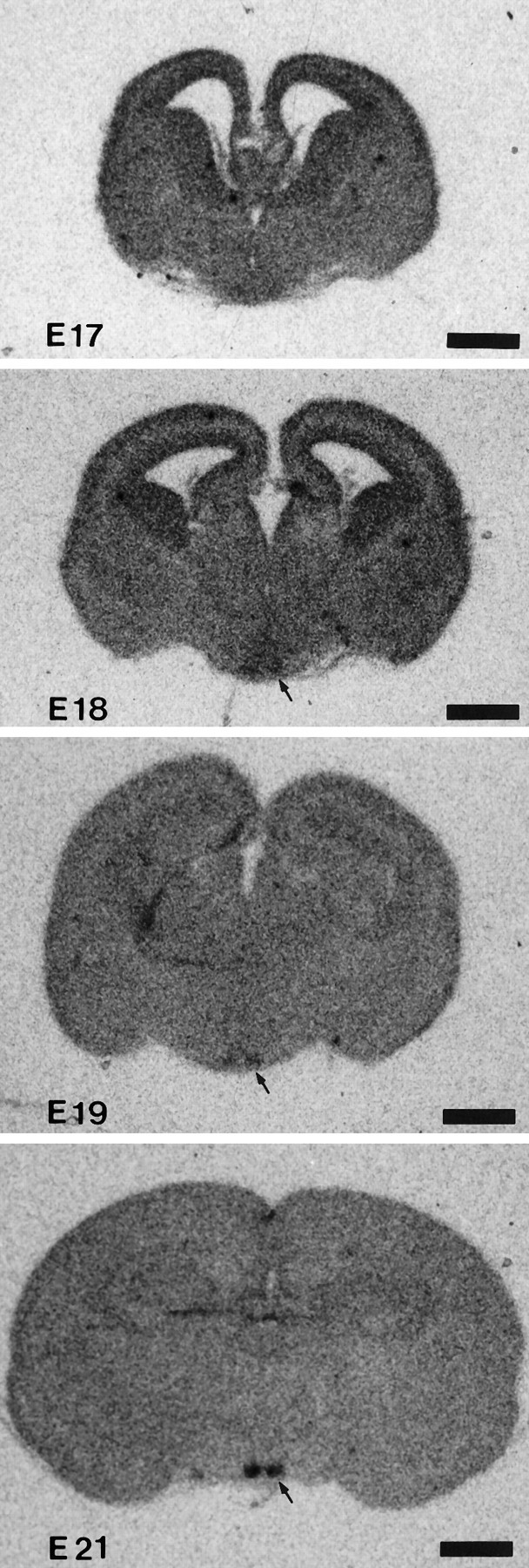
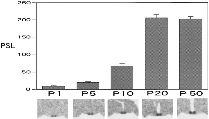
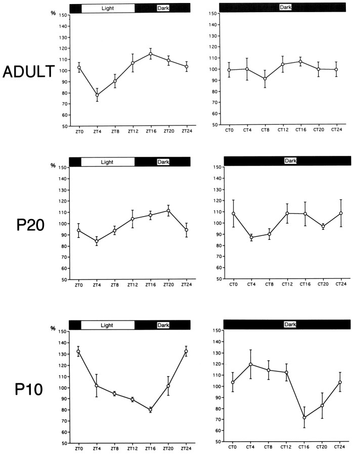
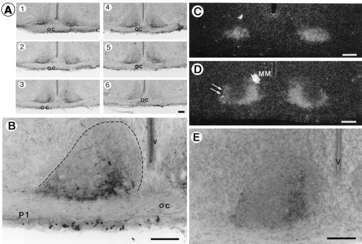
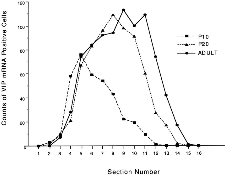
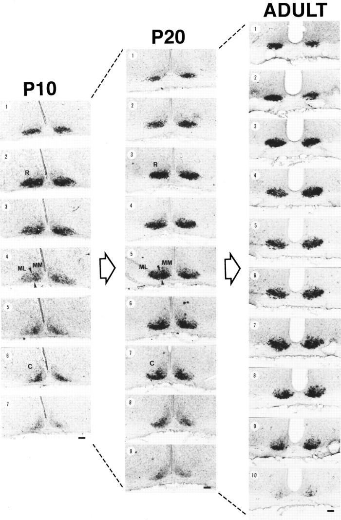
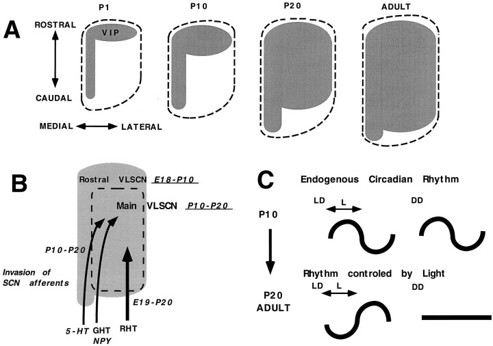
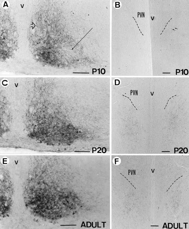
Development of the daily rhythm of vasoactive intestinal peptide (VIP) mRNA in the rat suprachiasmatic nucleus (SCN), a main locus of circadian oscillation, was investigated by in situ hybridization. The phenotypic expression of VIP neurons occurred in two developmental stages in the ventrolateral portion of the SCN (VLSCN): the first was found before birth in the rostral part, and the second occurred in the main part between postnatal day (P) 10 and P20. The latter period coincided with the time that the massive VIP-efferent fibers project to the subparaventricular zone. In the adult and P20, the VIP mRNA signals of the SCN showed a clear diurnal rhythm with a trough in the light phase and a peak in the dark phase under light/dark (LD) conditions, but under constant dark (DD) conditions, no VIP mRNA fluctuations were observed. At P10, however, it was found that SCN VIP mRNA showed a peak at the transition from night to day and a trough at early dark period in LD conditions, in sharp contrast to the night peak in the adult rhythm. In DD conditions, a light-phase peak and a dark-phase trough were also observed at P10, contrasting the arrhythmic feature at adult stage. The present findings suggest that daily VIP rhythm was first generated in the early developed clock-controlled rostral SCN neurons, and later regulated by light-dependent main VLSCN neurons.


Figures

Fig. 1.
Fig. 1.
In situ hybridization of VIP mRNA in the prenatal period. Photomicrographs were taken from β-max film. Scale bar, 1 mm.
Fig. 2.
Fig. 2.
Postnatal development of VIP mRNA levels shown as the total amount of PSL on imaging plates. The data are shown as the mean ± SEM, and examples of the images are shown under the time schedule.
Fig. 3.
Fig. 3.
Diurnal and circadian profiles of VIP mRNA levels in the SCN at P10, P20, and adult (P50).
Fig. 4.
Fig. 4.
Immunocytochemistry (A, B) and emulsion images of in situ hybridization (C–E) of VIP at P1. A, Rostrocaudal arrangement of VIP immunoreactivity in the SCN. Note that immunoreactivities were distributed not in the whole part, but only in limited areas of the VLSCN in most sections. B, Higher magnification photomicrograph of A3. Boundaries of SCN at P1 are encircled with dotted lines. C, D, Dark-field photomicrographs of VIP mRNA at the most rostral (C) and the middle (D) level of the SCN. At the middle level of the VLSCN, the signal mass was detected in the main medial (MM) part (thick short arrow), extending ventral thin wings to the lateral part (thin long arrows). E, Bright-field higher magnification photomicrograph of D counterstained with cresyl violet. oc, Optic chiasma; v, third ventricle. Scale bars, 100 μm.
Fig. 6.
Fig. 6.
Counts of VIP mRNA-positive cells in each rostrocaudally arranged section in one representative rat at each stage (P10, P20, and adult). Section 1 was designated as the most rostral SCN section recognized by Nissl staining, and the section number was sequentially ordered at the rostrocaudal direction. Section thickness was 40 μm at all stages.
Fig. 5.
Fig. 5.
Emulsion images of VIP mRNA in the SCN at P10, P20, and adult (P50). All serial sections (40 μm thickness) from one rat at each stage are presented in order of rostrocaudal direction. At middle-level sections (P10, number 4; P20, number5) of P10 and P20, VLSCN was subdivided further into lateral (middle-lateral; ML) and medial (middle-medial;MM) parts, and the borders of MM and ML are presented as arrowheads. (R) and (C) are representative sections of rostral and caudal SCN, respectively. Scale bars, 100μm.
Fig. 8.
Fig. 8.
A, Schematic representation of the development of VIP mRNA at P1, P10, P20, and adult in the SCN in thehorizontal plane. Gray area represents the VIP cell body distribution, and the dotted lineshows the border of SCN outlined by Nissl staining. B, Schema of the phenotypic expression time (underlined) in each compartment of the SCN, and the invasion time of SCN afferents [serotonin (5-HT), GHT-NPY, andRHT]. Invasion time of the SCN afferents was adapted from Takatsuji et al. (1995). C, Schema of the developmental change of VIP mRNA rhythms. See Discussion for details.
Fig. 7.
Fig. 7.
Immunocytochemistry of VIP at P10 (A, B), P20 (C, D), and adult (P50) (E, F) in SCN (A, C, E) and the subparaventricular zone (B, D, F). A long thin arrow indicates the main portion of the VLSCN, and athick hollow arrow indicates the dorsomedial part of the SCN. PVN, Hypothalamic paraventricular nucleus;v, third ventricle. Scale bars, 100 μm.

References

    1. Albers HE, Minamitani N, Stopa E, Ferris CF. Light selectively alters vasoactive intestinal peptide and peptide histidine isoleucine immunoreactivity within the rat suprachiasmatic nucleus. Brain Res. 1987;437:189–192. - PubMed
    1. Albers HE, Stopa EG, Zoeller RT, Kauer JS, King JC, Fink JS, Mobtaker H, Wolfe H. Day-night variation in prepro vasoactive intestinal peptide/peptide histidine isoleucine mRNA within the rat suprachiasmatic nucleus. Mol Brain Res. 1990;7:85–89. - PubMed
    1. Altman J, Bayer SA. Development of the diencephalon in the rat. I. Autoradiographic study of the time of origin and settling pattern of neurons of the hypothalamus. J Comp Neurol. 1978;182:945–972. - PubMed
    1. Amemiya Y, Miyahara J. Imaging plate illuminates many fields. Nature. 1988;336:89–90. - PubMed
    1. Anderson DJ. The neural crest cell lineage problem: neuropoiesis? Neuron. 1989;3:1–12. - PubMed

Publication types

Substances