

H. M.'s medial temporal lobe lesion: findings from magnetic resonance imaging
- PMID: 9133414
- PMCID: PMC6573687
- DOI: 10.1523/JNEUROSCI.17-10-03964.1997
H. M.'s medial temporal lobe lesion: findings from magnetic resonance imaging
Abstract
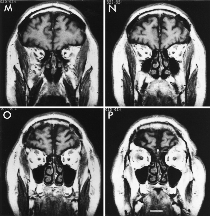
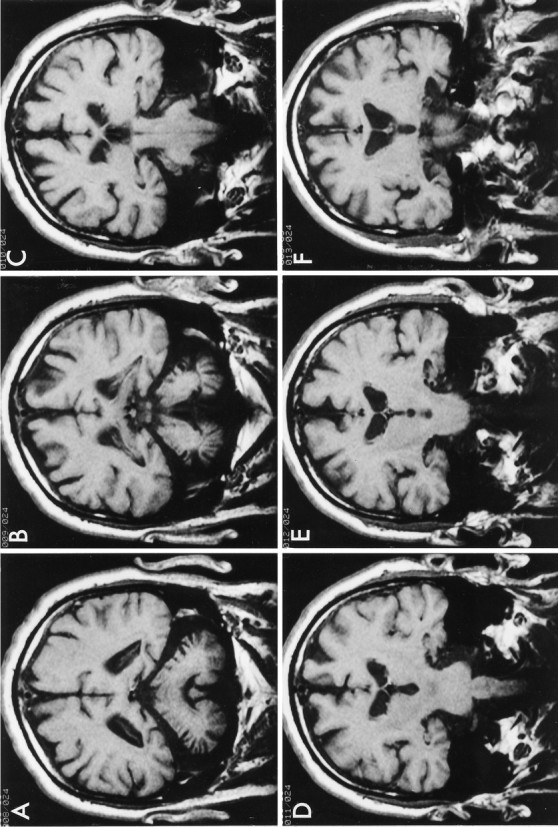
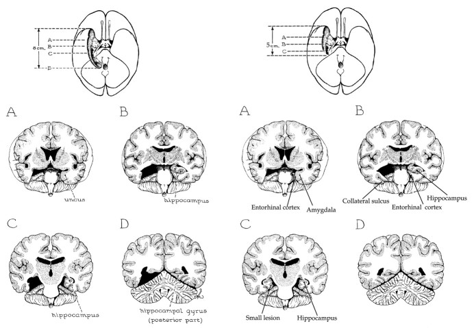
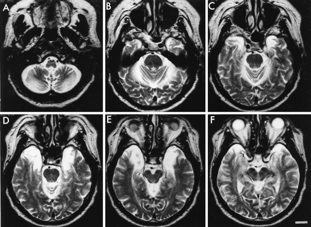
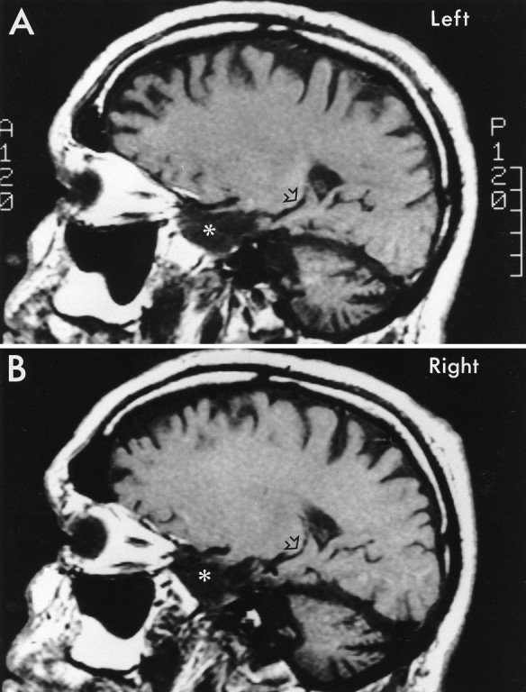
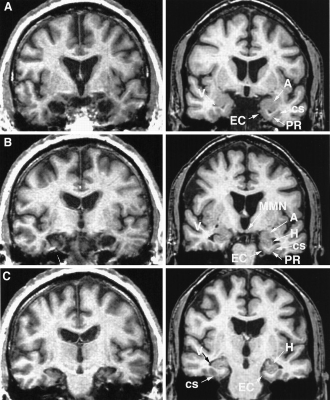
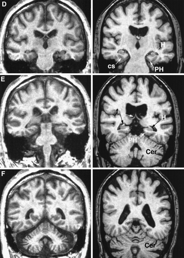
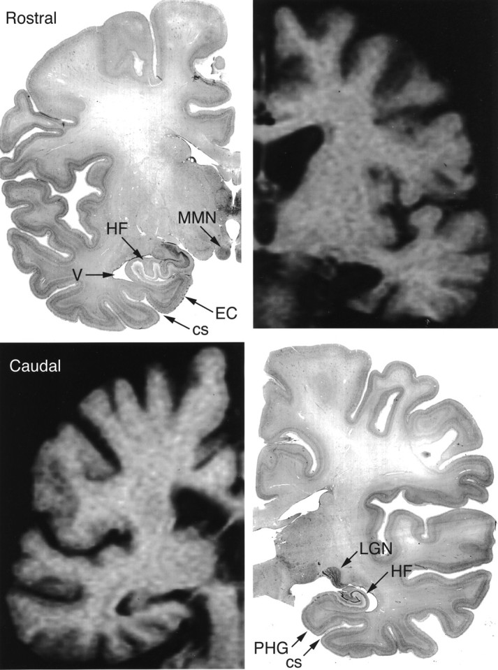
Although neuropsychological studies of the amnesic patient H. M. provide compelling evidence that normal memory function depends on the medial temporal lobe, the full extent of his surgical resection has not been elucidated. We conducted magnetic resonance imaging studies to specify precisely the extent of his bilateral resection and to document any other brain abnormalities. The MRI studies indicated that the lesion was bilaterally symmetrical and included the medial temporal polar cortex, most of the amygdaloid complex, most or all of the entorhinal cortex, and approximately half of the rostrocaudal extent of the intraventricular portion of the hippocampal formation (dentate gyrus, hippocampus, and subicular complex). The collateral sulcus was visible throughout much of the temporal lobe, indicating that portions of the ventral perirhinal cortex, located on the banks of the sulcus, were spared; the parahippocampal cortex (areas TF and TH) was largely intact. The rostrocaudal extent of the ablation was approximately 5.4 cm (left) and 5.1 cm (right). The caudal 2 cm, approximately, of the hippocampus body (normal length, approximately 4 cm) was intact, although atrophic. The temporal stem was intact. Outside the temporal lobes, the cerebellum demonstrated marked atrophy, and the mammillary nuclei were shrunken. The lateral temporal, frontal, parietal, and occipital lobe cortices appeared normal for age 66 years. The mediodorsal thalamic nuclei showed no obvious radiological changes. These findings reinforce the view that lesions of the hippocampal formation and adjacent cortical structures can produce global and enduring amnesia and can exacerbate amnesia beyond that seen after more selective hippocampal lesions.
Figures

References
-
- Alonso JR, Amaral DG. Cholinergic innervation of the primate hippocampal formation. I. Distribution of choline acetyltransferase immunoreactivity in the Macaca fascicularis and Macaca mulatta monkeys. J Comp Neurol. 1995;355:135–170. - PubMed
-
- Amaral DG, Insausti R. The hippocampal formation. In: Paxinos G, editor. The human nervous system. Academic; San Diego: 1990. pp. 711–755.
-
- Amaral DG, Insausti R, Cowan WM. The entorhinal cortex of the monkey. I. Cytoarchitectonic organization. J Comp Neurol. 1987;264:326–355. - PubMed
-
- Beason-Held L, Rosene DL, Moss MB. Memory deficits associated with ibotenic acid lesions of the hippocampal formation in rhesus monkeys. Soc Neurosci Abstr. 1993;19:438.
Publication types
MeSH terms
Grants and funding
LinkOut - more resources
Full Text Sources
Miscellaneous