Clinical and molecular genetic characterisation of a family segregating autosomal dominant retinitis pigmentosa and sensorineural deafness
- PMID: 9135384
- PMCID: PMC1722127
- DOI: 10.1136/bjo.81.3.207
Clinical and molecular genetic characterisation of a family segregating autosomal dominant retinitis pigmentosa and sensorineural deafness
Abstract
Aims/background: To characterise clinically a large kindred segregating retinitis pigmentosa and sensorineural hearing impairment in an autosomal dominant pattern and perform genetic linkage studies in this family. Extensive linkage analysis in this family had previously excluded the majority of loci shown to be involved in the aetiologies of RP, some other forms of inherited retinal degeneration, and inherited deafness.
Methods: Members of the family were subjected to detailed ophthalmic and audiological assessment. In addition, some family members underwent skeletal muscle biopsy, electromyography, and electrocardiography. Linkage analysis using anonymous microsatellite markers was performed on DNA samples from all living members of the pedigree.
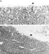
Results: Patients in this kindred have a retinopathy typical of retinitis pigmentosa in addition to a hearing impairment. Those members of the pedigree examined demonstrated a subclinical myopathy, as evidence by abnormal skeletal muscle histology, electromyography, and electrocardiography. LOD scores of Zmax = 3.75 (theta = 0.10), Zmax = 3.41 (theta = 0.10), and Zmax = 3.25 (theta = 0.15) respectively were obtained with the markers D9S118, D9S121, and ASS, located on chromosome 9q34-qter, suggesting that the causative gene in this family may lie on the long arm (q) of chromosome 9.
Conclusions: These data indicate that the gene responsible for the phenotype in this kindred is located on chromosome 9 q. These data, together with evidence that a murine deafness gene is located in a syntenic area of the mouse genome, should direct the research community to consider this area as a candidate region for retinopathy and/or deafness genes.
Figures


References
Publication types
MeSH terms
Substances
Grants and funding
LinkOut - more resources
Full Text Sources
Medical
Miscellaneous
