Classical Article
. 1997 Feb;120(4 Suppl):337-57; discussion 334-6.
doi: 10.1111/j.1476-5381.1997.tb06815.x.

Evidence that adenosine triphosphate or a related nucleotide is the transmitter substance released by non-adrenergic inhibitory nerves in the gut. 1970

Classical Article

Evidence that adenosine triphosphate or a related nucleotide is the transmitter substance released by non-adrenergic inhibitory nerves in the gut. 1970

G Burnstock et al. Br J Pharmacol. 1997 Feb.

Abstract

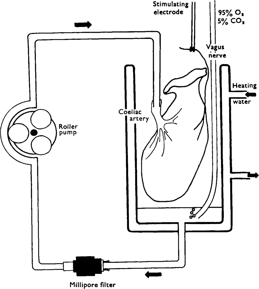
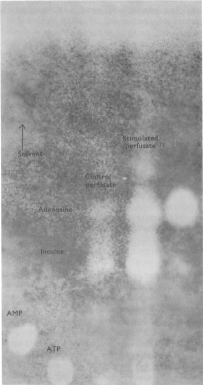
  1. Stimulation of the vagal non-adrenergic inhibitory innervation caused the release of adenosine and inosine into vascular perfusates from the stomachs of guinea-pigs and toads.

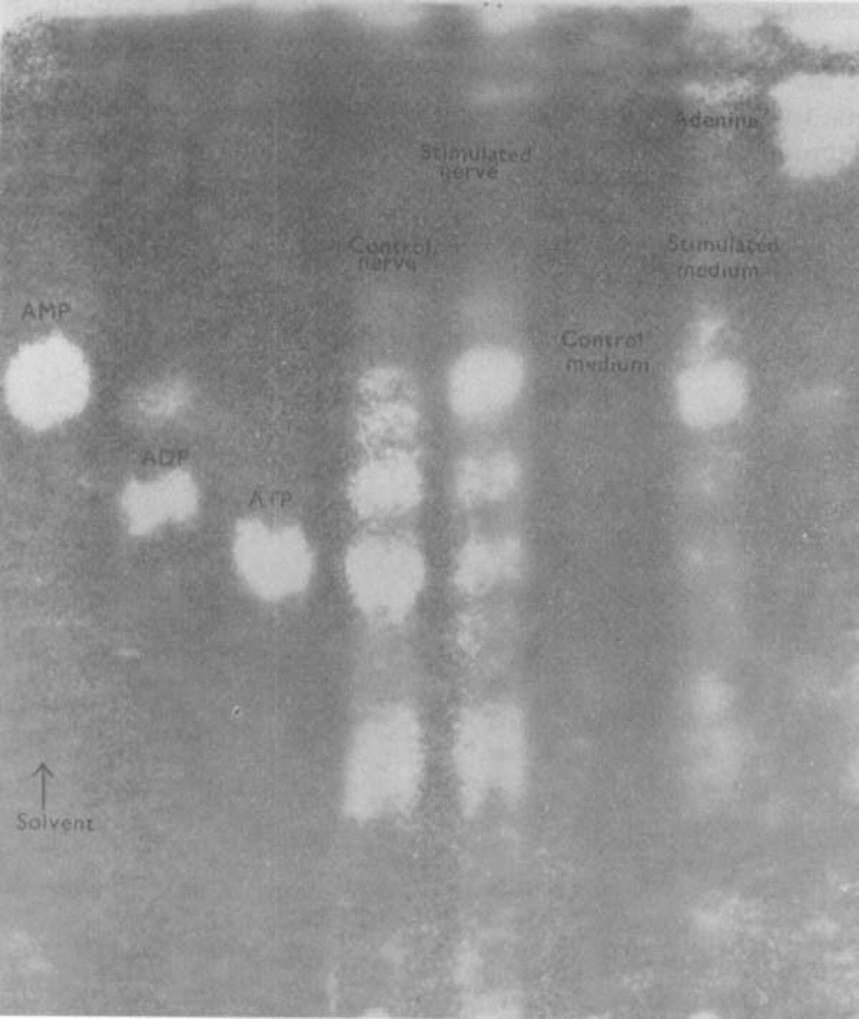
  2. Stimulation of portions of Auerbach's plexus isolated from turkey gizzard caused the release of adenosine triphosphate (ATP), adenosine diphosphate (ADP) and adenosine monophosphate (AMP).

  3. ATP, added to solutions perfused through the toad stomach vasculature, was broken down to adenosine, inosine and adenine.

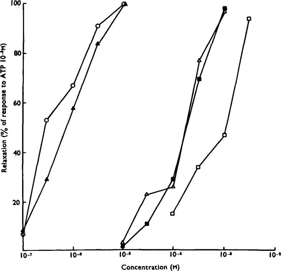
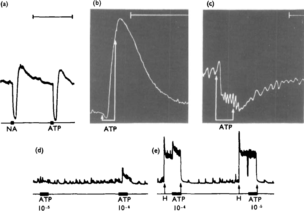
  4. Of a series of purine and pyrimidine derivatives tested for inhibitory activity on the guinea-pig isolated taenia coli, ATP and ADP were the most potent.

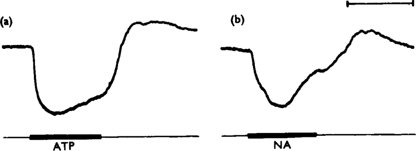
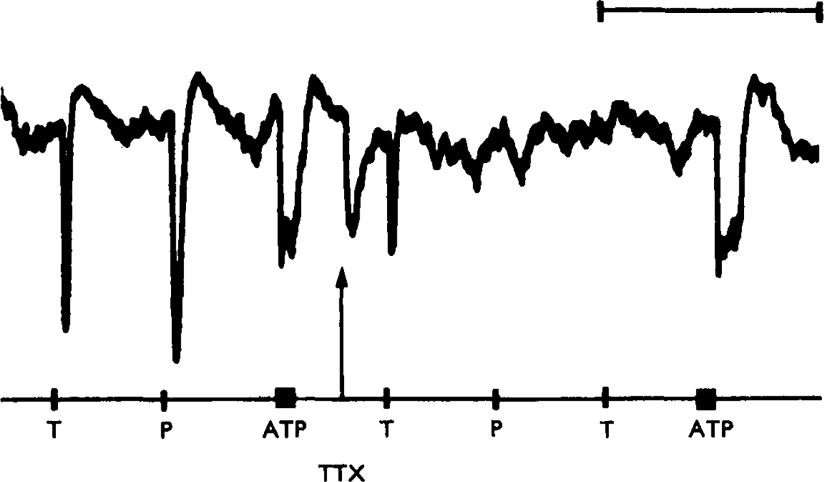
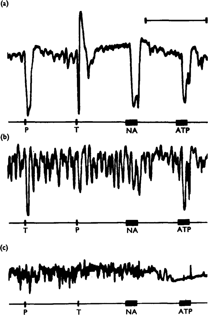
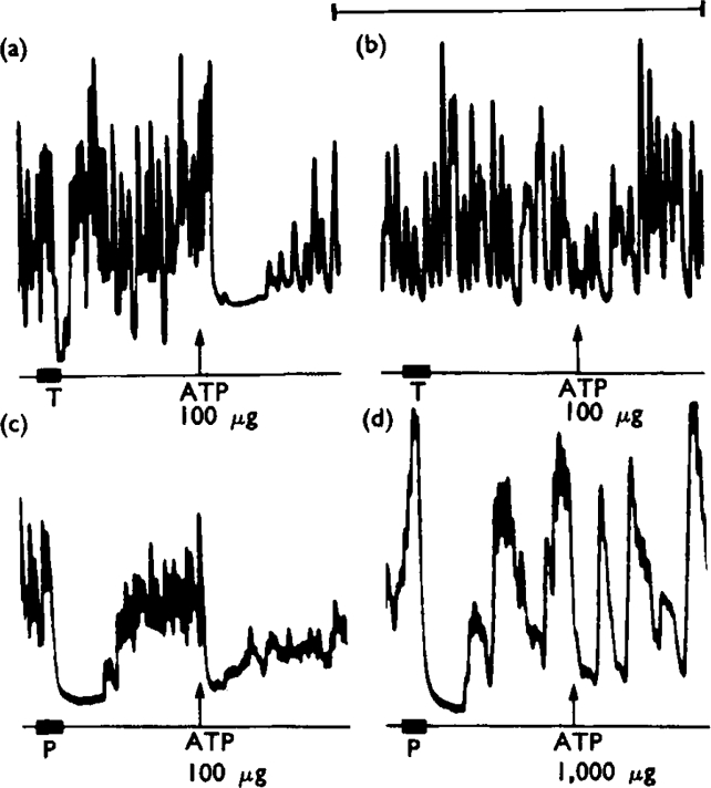
  5. ATP caused inhibition of twelve other gut preparations previously shown to contain non-adrenergic inhibitory nerves. The inhibitory action of ATP was not prevented by tetrodotoxin.

  6. Quinidine antagonized relaxations of the guinea-pig taenia coli caused by catecholamines or adrenergic nerve stimulation. Higher concentrations of quinidine antagonized relaxations caused by ATP or non-adrenergic inhibitory nerve stimulation.

  7. When tachyphylaxis to ATP had been produced in the rabbit ileum, there was a consistent depression of the responses to non-adrenergic inhibitory nerve stimulation but not of responses to adrenergic nerve stimulation.

  8. It is suggested that ATP or a related nucleotide is the transmitter substance released by the non-adrenergic inhibitory innervation of the gut.


Figures

Fig. 1
Fig. 1
Diagram of apparatus used for closed-circuit perfusion of stomach preparations. A small volume (2·5 ml) of nutrient solution is pumped by the roller pump through a cannula into the coeliac artery. Fluid draining from the gastric veins collects in the bottom of the constant temperature organ bath where it is bubbled with 95% oxygen and 5% carbon dioxide. The fluid is then drawn through a Millipore filter to be recycled. Shielded platinum wire ring-electrodes are placed around the vagus nerves for stimulation
Fig. 2
Fig. 2
Chromatographic evidence for purine release from toad stomach stimulated via the vagosympathetic nerve. The figure shows an ultraviolet photoprint of a chromatogram spotted with (from the left) AMP, ATP, perfusate from unstimulated stomach, perfusate from stimulated stomach, adenosine. Chromatogram run for 12 h ascending in solvent 1 (see text). Vagosympathetic nerve trunks were stimulated for 45 s in every 2 min for a total of 30 min with pulses of 2 ms duration, at 30 Hz; the voltage was progressively increased from 5 to 40 V. Note that stimulation increases the amounts of adenosine and inosine released from the stomach.
Fig. 3
Fig. 3
Release of nucleotides from isolated Auerbach's plexus of turkey gizzard. The figure shows an ultraviolet photoprint of a chromatogram spotted with, from the left, AMP, ADP, ATP, extracted unstimulated plexus, extracted stimulated plexus, medium of unstimulated plexus, medium of stimulated plexus (pulses of 2 ms at 50 Hz, trains of 45 s every 2 min for 30 min, voltage progressively increasing from 5 to 50 V), adenine. Note that stimulation causes the release of AMP and traces of ADP and ATP into the medium. Comparable levels of nucleotides were found in extracts of stimulated and unstimulated plexuses. Chromatogram run for 12 h ascending in solvent 2 (see text)
Fig. 4
Fig. 4
Breakdown of ATP perfused through the toad stomach vascular bed. The figure shows an ultraviolet photoprint of a chromatogram spotted with (from the left) AMP, ATP + ADP control perfusate, perfusate with ATP added before recycling, adenosine, inosine, IMP, adenine. Chromatogram run for 12 h ascending in solvent 1. Note that the ATP added to the medium was broken down to adenosine and inosine
Fig. 5
Fig. 5
Relative potencies of purine compounds in causing relaxation of the isolated guinea-pig taenia coli. Ordinate: amplitude of relaxation as a percentage of the response to ATP (10−5m). Abscissa: molar concentration (log scale). Dose-response curves are shown for ATP (▴), ADP (○), AMP (δ), adenosine (▪) and GMP (□). The tissues were exposed to the agonists for 30 s. Each point is the mean of values obtained on three preparations
Fig. 6
Fig. 6
Relative rapidity of gut responses to ATP. Consecutive, approximately equal sub-maximal relaxations of a guinea-pig isolated taenia coli, (a) caused by ATP (10−5 g/ml) and (b) by noradrenaline (NA, 10−7 g/ml). Drugs were applied for 1 mm, as shown by the bars. Note that the ATP response reaches its maximum after 19 s whereas the response to noradrenaline takes 28 s to become maximal. Time marker, 1 minute
Fig. 7
Fig. 7
Isolated guinea-pig taenia coli treated with hyoscine (4× 10⁻⁷ g/ml). Control responses to transmural stimulation (T, 10 Hz for 10 s), perivascular nerve stimulation (P, 10 Hz for 10 s) and ATP (bar, 2× 10⁻⁶ g/ml) are shown at the left of the trace. At the arrow, tetrodotoxin (TTX, 1·5 ×10−7 g/ml) was added to the organ bath. Responses to both transmural and perivascular nerve stimulation were rapidly abolished, but the response to ATP was not reduced. Time marker, 10 minutes
Fig. 8
Fig. 8
Responses of selected gut segments to ATP. Preparations treated with hyoscine (10−6 g/ml). (a), Guinea-pig isolated taenia coli. Both noradrenaline (NA, 2 × 10⁻⁷ g/ml) and ATP (ATP, 10−5 g/ml), applied for 15 s (bars) caused relaxation, followed by a secondary “rebound” contraction when the drugs were washed from the organ bath. (b), Isolated rat gastric fundus strip. ATP (10−6 g/ml), applied for 1 min between arrows, caused an initial slight relaxation followed by a contraction while the drug was still in the organ bath. (c). Isolated, Ringer-perfused toad stomach. ATP (2·5 × 10−7 g/ml in perfusate), applied for 6 min between the arrows, caused a slight initial rise followed by a marked fall in intragastric pressure. (d), (e), Guinea-pig isolated ileum. In a low-tone preparation (d) ATP (10−5 g ml) applied for 60 s (bar) was without effect. A higher concentration (10−4 g/ml) caused contraction. (e), When the preparation was contracted with histamine (H. 6·5 × 10−8 g/ml, applied between arrows), ATP 10−4 g/ml still caused contraction but 10−3 g/ml caused a brief relaxation. Time markers, 5 minutes. The time marker for (a) also refers to (d) and (c)
Fig. 9
Fig. 9
Effects of quinidine on inhibitory responses of the guinea-pig taenia coli, treated with hyoscine (10−6 g/ml). Panel (a) shows control responses to perivascular nerve stimulation (P, 20 Hz for 10 s), transmural stimulation (T. 10 Hz for 10 s), noradrenaline (NA. 5 × 10⁻⁸ g/ml for 40 s) and ATP (ATP. 4 × 10⁻⁵ g/ml for 40 s). In (b). the preparation had been treated with quinidine (5 × 10⁻⁵ g/ml) for 65 minutes. The responses to perivascular nerve stimulation and to noradrenaline were virtually abolished, whereas the responses to transmural stimulation and to ATP were not reduced in amplitude. In (c), the preparation had been treated with quinidine (2 × 10⁻¹ g/ml) for a further 20 minutes. The responses to transmural stimulation and to ATP were now also abolished. Time marker, 5 minutes
Fig. 10
Fig. 10
Effects of desensitization of rabbit isolated ileum to ATP. Preparations treated with hyoscine (10−6 g/ml). Panels (a) and (b) show responses of a preparation subjected to degenerative section of the perivascular adrenergic innervation 6 days previously. In (a), transmural stimulation (T, 10 Hz for 20 s) caused inhibition. At the arrow, ATP (100 μg to make 10−5 g/ml) added to the organ bath and left in contact with the tissue caused a transient inhibition. Panel (b) starts 15 min after (a). In the interval, three further applications of ATP (100 μg) had been made. The responses to both transmural stimulation and ATP (100 μg) are virtually abolished. Calcium concentration of medium four times normal. Panels (c) and (d) show responses of a preparation with normal perivascular adrenergic innervation. In (c), perivascular nerve stimulation (P, 10 Hz for 20 s) and ATP (100 μg to make 10−5 g/ml, added at arrow and left in) caused inhibition. Panel (d) starts 40 min after (c), after five 100 μg and two 1,000 μg applications of ATP. ATP (1,000 μg applied at the arrow) was ineffective but the response to perivascular nerve stimulation was still present. Calcium concentration of medium twice normal. Time marker, 5 minutes

Similar articles

Cited by

References

    1. Abood LG, Koketsu K, Miyamoto S. Outflux of various phosphates during membrane depolarization of excitable tissue. Am. J. Physiol. 1962;202:469–474. - PubMed
    1. Ambache N, Freeman MA. Atropine-resistant longitudinal muscle spasms due to excitation of non-cholinergic neurones in Auerbach's plexus. J. Physiol., Lond. 1968;199:705–728. - PMC - PubMed
    1. Axelsson J, Holmberg B. The effects of extracellularly applied ATP and related compounds on electrical and mechanical activity of the smooth muscle taenia coli from the guinea-pig. Acta physiol. scand. 1969;75:149–156. - PubMed
    1. Barsoum GS, Gaddum JH. The pharmacological estimation of adenosine and histamine in blood. J. Physiol., Lond. 1935;85:1–14. - PMC - PubMed
    1. Bell C. Differential effects of tetrodotoxin on sympathomimetic actions of nicotine and tyramine. Br. J. Pharmac. Chemother. 1968;32:96–103. - PMC - PubMed

MeSH terms

Substances

Personal name as subject

LinkOut - more resources