

doi: 10.1073/pnas.94.10.4937.
In vitro selection and evolution of functional proteins by using ribosome display
Affiliations
- PMID: 9144168
- PMCID: PMC24609
- DOI: 10.1073/pnas.94.10.4937
Item in Clipboard
In vitro selection and evolution of functional proteins by using ribosome display
Proc Natl Acad Sci U S A.
.
Abstract
We report here a system with which a correctly folded complete protein and its encoding mRNA both remain attached to the ribosome and can be enriched for the ligand-binding properties of the native protein. We have selected a single-chain fragment (scFv) of an antibody 10(8)-fold by five cycles of transcription, translation, antigen-affinity selection, and PCR. The selected scFv fragments all mutated in vitro by acquiring up to four unrelated amino acid exchanges over the five generations, but they remained fully compatible with antigen binding. Libraries of native folded proteins can now be screened and made to evolve in a cell-free system without any transformation or constraints imposed by the host cell.
Figures
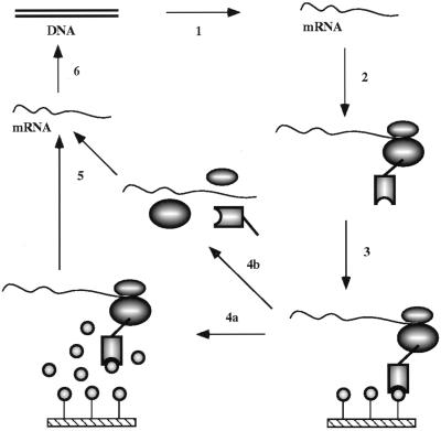
Principle of in vitro ribosome display for screening native protein (scFv) libraries for ligand (antigen) binding. Step 1, a DNA scFv library is first amplified by PCR, whereby a T7 promoter, ribosome-binding site, and stem–loops are introduced, and then transcribed to RNA. Step 2, after purification, mRNA is translated in vitro in an Escherichia coli S-30 system in the presence of different factors enhancing the stability of ribosomal complexes and improving the folding of the scFv antibodies on the ribosomes. Translation is stopped by cooling on ice, and the ribosome complexes are stabilized by increasing the magnesium concentration. Step 3, the desired ribosome complexes are affinity selected from the translation mixture by binding of the native scFv to the immobilized antigen. Unspecific ribosome complexes are removed by intensive washing. Step 4, the bound ribosome complexes can then be dissociated by EDTA (step 4b), or whole complexes can be specifically eluted with antigen (step 4a). Step 5, RNA is isolated from the complexes. Step 6, isolated mRNA is reverse transcribed to cDNA, and cDNA is then amplified by PCR. This DNA is then used for the next cycle of enrichment, and a portion can be analyzed by cloning and sequencing and/or by ELISA or RIA.
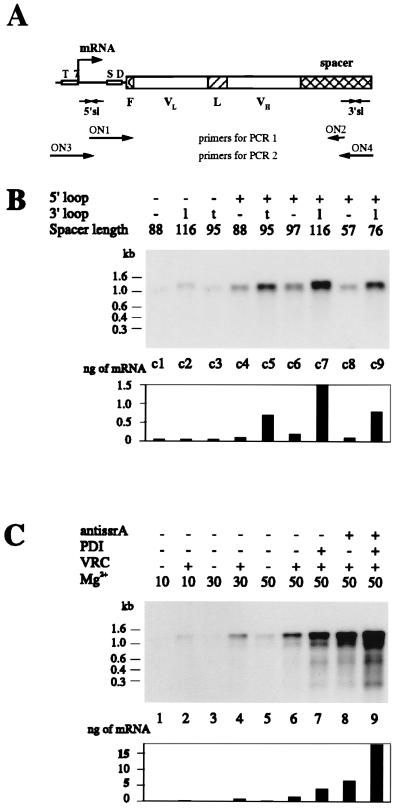
(A) Schematic drawing of the scFv construct used for ribosome display. T7 denotes the T7 promoter, SD the ribosome-binding site, F the 3-amino-acid-long FLAG (28) with N-terminal methionine, VL and VH the variable domains of the scFv fragment, L the linker, spacer the part of protein construct connecting the folded scFv to the ribosome, and 5′sl and 3′sl the stem–loops on the 5′ and 3′ ends of the mRNA. The arrow indicates the transcriptional start. The strategy for the PCR amplification is shown. (B) The amount of the scFvhag mRNA bound to the polysome complexes is influenced by the secondary structure of its ends and the length of the spacer connecting the folded scFv to the ribosome. Different constructs of scFvhag mRNA were used for one cycle of ribosome display (constructs c1–c9). None of them contained a stop codon. Each species was tested separately. After affinity selection of the ribosome complexes from 100 μl of translation mixtures, mRNAs were isolated and analyzed by Northern hybridization. The presence of the 5′ stem–loop is labeled + or −; that of the 3′ stem–loop is labeled as − when absent, l when derived from lpp term, or t when derived from T3Te. The spacer length indicates the number of amino acids following the scFv and connecting it to the ribosome, including the translated stem–loop. The lengths of RNA in kb are derived from RNA molecular weight marker III (Boehringer Mannheim). The bar graph shows the quantified amounts of mRNA from fluoroimager analysis. (C) Effect of additives on the amount of mRNA bound to the ribosome complexes. The mRNA of the scFvhag construct c7 was used for one cycle of ribosome display. Samples in B lane c7 and C lane 6 are identical. When indicated, 3.5 mM anti-ssrA oligonucleotide ON10 (5′-TTAAGCTGCTAAAGCGTAGTTTTCGTCGTTTGCGACTA-3′), 35 mg/ml protein disulfide isomerase (PDI), or 1 mg/ml VRC was included in the translation mixture. Translations were stopped by addition of Mg(OAc)2 to the final concentration indicated (in mM) and by cooling on ice. After affinity selection of the ribosome complexes from 100 μl of translation mixtures, mRNAs were isolated and analyzed by Northern hybridization. The bar graph shows the quantified amounts of mRNA from fluoroimager analysis.
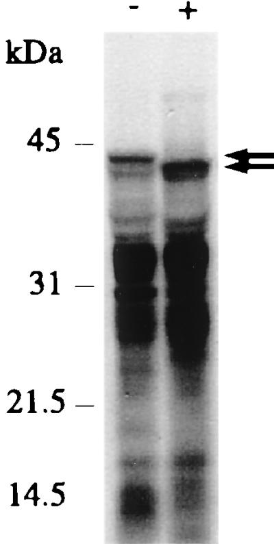
Anti-ssrA antisense oligonucleotide ON10 (Fig. 2C) decreases the molecular mass of the longest protein species (arrows). In vitro translation was performed using [35S]methionine and scFvhag c7 mRNA. Reactions were carried out in the absence (−) or presence (+) of 3.5 mM oligonucleotide ON10. An SDS/PAGE of translation products is shown.
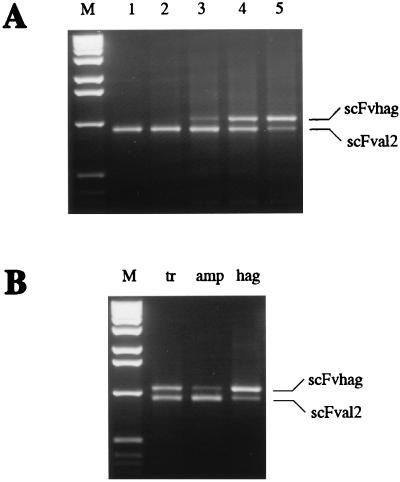
(A) Enrichment of the scFvhag ribosome complexes from mixtures with scFvAL2 by ribosome display. mRNA of scFvhag c5 was diluted 108 times with mRNA of scFvAL2, and the mixture was used for ribosome display. After affinity selection of scFvhag ribosome complexes, mRNA was isolated and reverse transcribed to cDNA using oligonucleotide ON5, and cDNA was amplified by PCR using oligonucleotides ON3 and ON7 and analyzed by agarose electrophoresis. Lanes 1–5 are PCR products amplified after the first to fifth cycles of the ribosome display, respectively. Lane M is the 1-kb DNA ladder (GIBCO/BRL) used as molecular weight markers. PCR products corresponding to scFvhag and scFvAL2 are labeled. (B) Enrichment of either scFvhag c5 or scFvAL2 ribosome complexes by ribosome display as a function of immobilized antigen. mRNAs of scFvhag and scFvAL2 were mixed in a 1:1 ratio and used for one cycle of ribosome display. After affinity selection, mRNA was isolated, reverse transcribed, amplified by PCR, and analyzed by agarose electrophoresis as in A. The same 1:1 mixture was affinity selected on immobilized transferrin (tr) as a control, ampicillin-transferrin conjugate (amp), or hemagglutinin-peptide-transferrin conjugate (hag). PCR products corresponding to scFvhag and scFvAL2 are labeled.
References
-
- Saffhill R, Schneider-Bernloehr H, Orgel L E, Spiegelman S. J Mol Biol. 1970;51:531–539. - PubMed
-
- Gold L, Polisky B, Uhlenbeck O, Yarus M. Annu Rev Biochem. 1995;64:763–797. - PubMed
-
- Irvine D, Tuerk C, Gold L. J Mol Biol. 1991;222:739–761. - PubMed
-
- Dower W J, Cwirla S E. In: Guide to Electroporation and Electrofusion. Chang D C, Chassy B M, Saunders J A, Sowers A E, editors. San Diego: Academic; 1992. pp. 291–301.
Publication types
MeSH terms
Substances
LinkOut - more resources
Full Text Sources
Other Literature Sources
