. 1997 May 13;94(10):4966-71.
doi: 10.1073/pnas.94.10.4966.

Expression, stability, and membrane integration of truncation mutants of bovine rhodopsin

Affiliations

Expression, stability, and membrane integration of truncation mutants of bovine rhodopsin

J A Heymann et al. Proc Natl Acad Sci U S A. .

Abstract

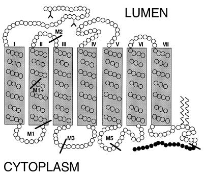
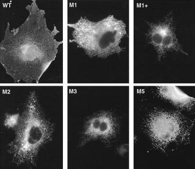
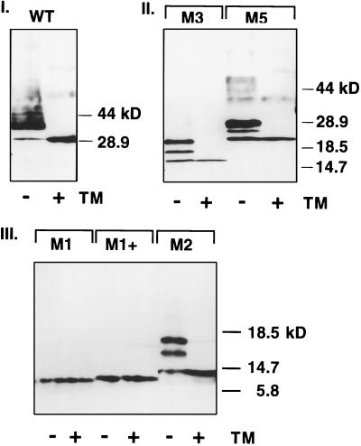
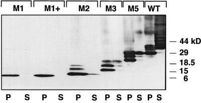
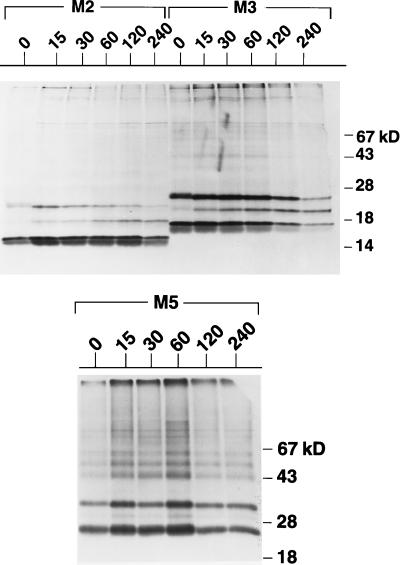
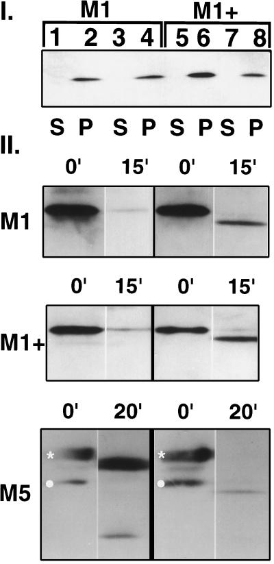
Premature termination of protein synthesis by nonsense mutations is at the molecular origin of a number of inherited disorders in the family of G protein-coupled seven-helix receptor proteins. To understand how such truncated polypeptides are processed by the cell, we have carried out COS-1 cell expression studies of mutants of bovine rhodopsin truncated at the first 1, 1.5, 2, 3, or 5 transmembrane segments (TMS) of the seven present in wild-type opsin. Our experiments show that successful completion of different stages in the cellular processing of the protein [membrane insertion, N-linked glycosylation, stability to proteolytic degradation, and transport from the endoplasmic reticulum (ER) membrane] requires progressively longer lengths of the polypeptide chain. Thus, none of the truncations affected the ability of the polypeptides to be integral membrane proteins. C-terminal truncations that generated polypeptides with fewer than two TMS resulted in misorientation and prevented glycosylation at the N terminus, whereas truncations that generated polypeptides with fewer than five TMS greatly destabilized the protein. However, all of the truncations prevented exit of the polypeptide from the ER. We conclude that during the biogenesis of rhodopsin, proper integration into the ER membrane occurs only after the synthesis of at least two TMS is completed. Synthesis of the next three TMS confers a gradual increase in stability, whereas the presence of more than five TMS is necessary for exit from the ER.


Figures

Figure 1
Figure 1
Secondary structural map of bovine opsin (21) showing the locations at which deletions were carried out. Sites of deletion are indicated by bars. Solid gray boxes represent the approximate boundaries of the TMS (I through VII) and the Y symbols indicate N-linked oligosaccharides. All mutants were tagged with the C-terminal 16 amino acids (black) of bovine opsin, which is the epitope recognized by the 1D4 monoclonal antibody. The lengths of the constructs, restriction sites within the bovine opsin gene (22) used to generate the mutant genes, and linker amino acid sequences introduced at the C-terminal portion of the protein preceding the 1D4 tag are: WT (348 a.a.), EcoRI–SalI, none; M5 (255 a.a.), EcoRI–PstI, AQQQE; M3 (157 a.a.), EcoRI–PvuI, ERYVVVCK; M2 (122 a.a.), EcoRI–NcoI, HGYFVFG; M1+ (103 a.a.), DLFMV; M1 (90 a.a.), EcoRI–HindIII, KLRTPLNY, respectively.
Figure 2
Figure 2
Immunolocalization of deletion mutants in transiently transfected COS-1 cells. Cells were fixed and permeabilized 18–20 hr after transfection and stained with the 1D4 antibody followed by labeling with a fluorescent secondary antibody.
Figure 3
Figure 3
Immunoblot analysis of wild-type opsin, I, of the M3 and M5 mutants, II, and of the M1, M1+, and M2 mutants III, transiently expressed in COS-1 cells in the absence (−) and presence (+) of tunicamycin (TM). DM (1% extracts of whole cells were used for wild-type opsin and the M2, M3, and M5 mutants. Because the M1 and M1+ mutants were not significantly extracted by 1% DM, the DM insoluble membrane pellet was used instead. Immunoblots were analyzed with the 1D4 antibody (24) which recognizes the C-terminal tag (I and II) or the B6–30N antibody (ref. ; III) which recognizes an epitope at the N terminus which includes at least one of the two consensus glycosylation sites, the N2 and N15.
Figure 4
Figure 4
Immunoblot analysis of pellet (P) and supernatant (S) fractions of DM extracts of microsomal membranes of COS-1 cells transiently expressing wild-type or mutant opsin polypeptides. Extraction was performed as described in Materials and Methods. Immunoblots were analyzed with the 1D4 antibody.
Figure 5
Figure 5
Pulse–chase labeling of the M2, M3, and M5 mutants. COS-1 cells transiently transfected with mutant opsins were labeled for 5 min with [35S]methionine and [35S]cysteine and incubated in unlabeled methionine and cysteine for indicated chase time in minutes above each lane. Opsin was immunoprecipitated with the 1D4 antibody, resolved by SDS/PAGE (10% for M5; 12.5% for M2, and M3), and visualized by fluorography.
Figure 6
Figure 6
I. Immunoblot showing pellet and supernatant fractions following treatment of microsomal membranes isolated from COS-1 cells transiently expressing the M1 (lanes 1–4) and M1+ (lanes 5–8) mutants with either 4 M urea or sodium carbonate at pH 11.5. For urea-treated samples, supernatant fractions are in lanes 1 and 5, and pellet fractions are in lanes 2 and 6. For sodium carbonate-treated samples, supernatant fractions are in lanes 3 and 7, and pellet fractions are in lanes 4 and 8. II. Trypsin treatment of mutants M1, M1+, and M5 in microsomes prepared from transiently transfected COS-1 cells. The immunoblots were probed with monoclonal antibodies specific to either the N terminus (B6–30N, left side) or the C terminus (1D4, right side). The protease accessible side of the microsomal membrane preparation is equivalent to the cytoplasmic side of the ER membrane. The predominant species present at the end of the reaction in the M5 mutant is derived from proteolysis at the C terminus. In contrast, the predominant species in the M1 and M1+ mutants are derived from proteolysis at the N terminus of the polypeptide, indicating a reversed orientation in the membrane. The minor bands observed in the M5 mutant at 20 min have mobilities which are consistent with N- and C-terminal fragments derived from cleavage of the doubly glycosylated species (∗) in the loop region between TMS 1 and TMS 2, but may also be derived from cleavage of the unglycosylated species (○).

References

    1. Coughlin S R. Curr Opin Cell Biol. 1994;6:191–197. - PubMed
    1. Spiegel A M. Annu Rev Physiol. 1996;58:143–170. - PubMed
    1. Vanrhee A M, Jacobson K A. Drug Dev Res. 1996;37:1–38. - PMC - PubMed
    1. Pan Y, Metzenberg A, Das S, Jing B, Gitschier J. Nat Genet. 1992;2:103–106. - PubMed
    1. Jacobson S G, Kemp C M, Cideciyan A V, Macke J P, Sung C H, Nathans J. Invest Ophthalmol Visual Sci. 1994;35:2521–2534. - PubMed

Publication types

MeSH terms

LinkOut - more resources