. 1997 May 13;94(10):4994-9.
doi: 10.1073/pnas.94.10.4994.

DAD1, the defender against apoptotic cell death, is a subunit of the mammalian oligosaccharyltransferase

Affiliations

DAD1, the defender against apoptotic cell death, is a subunit of the mammalian oligosaccharyltransferase

D J Kelleher et al. Proc Natl Acad Sci U S A. .

Abstract

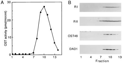
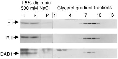
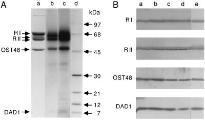
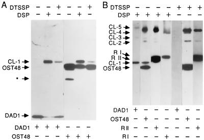
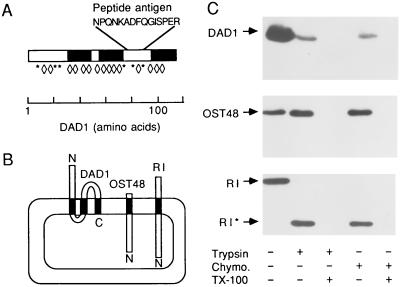
DAD1, the defender against apoptotic cell death, was initially identified as a negative regulator of programmed cell death in the BHK21-derived tsBN7 cell line. Of interest, the 12.5-kDa DAD1 protein is 40% identical in sequence to Ost2p, the 16-kDa subunit of the yeast oligosaccharyltransferase (OST). Although the latter observation suggests that DAD1 may be a mammalian OST subunit, biochemical evidence to support this hypothesis has not been reported. Previously, we showed that canine OST activity is associated with an oligomeric complex of ribophorin I, ribophorin II, and OST48. Here, we demonstrate that DAD1 is a tightly associated subunit of the OST both in the intact membrane and in the purified enzyme. Sedimentation velocity analyses of detergent-solubilized WI38 cells and canine rough microsomes show that DAD1 cosediments precisely with OST activity and with the ribophorins and OST48. Radioiodination of the purified OST reveals that DAD1 is present in roughly equimolar amounts relative to the other subunits. DAD1 can be crosslinked to OST48 in intact microsomes with dithiobis(succinimidylpropionate). Crosslinked ribophorin II-OST48 heterodimers, DAD1-ribophorin II-OST48 heterotrimers and DAD1-ribophorin I-ribophorin II-OST48 heterotetramers also were detected. The demonstration that DAD1 is a subunit of the OST suggests that induction of a cell death pathway upon loss of DAD1 in the tsBN7 cell line reflects the essential nature of N-linked glycosylation in eukaryotes.


Figures

Figure 1
Figure 1
Cosedimentation of DAD1 with OST activity, ribophorins I and II, and OST48. (A) A digitonin–high salt extract prepared from DK-RM was resolved by preparative glycerol density gradient centrifugation. Fraction 1 is the top of the gradient. Duplicate aliquots from each of 14 gradient fractions were assayed for OST activity. (B) Aliquots from each gradient fraction were analyzed on protein immunoblots using antibodies to ribophorin I (R I), ribophorin II (R II), OST48, and DAD1. The 64-kDa-glycosylated and 63-kDa-nonglycosylated forms of ribophorin II migrate as a doublet (13).
Figure 2
Figure 2
Total cellular DAD1 cosediments with the OST subunits. Intact WI38 fibroblasts were solubilized in digitonin–high salt solution as described. The total homogenate (T) was centrifuged for 15 min at 160,000 × g to prepare a detergent-solubilized supernatant (S) and a detergent-insoluble pellet (P). The digitonin–high salt supernatant (S) was resolved by glycerol density gradient centrifugation into 14 fractions. Fraction 1 is the top of the gradient. Samples corresponding to 3% of the total homogenate, 3% of the supernatant, and 6% of the pellet were resolved on one polyacrylamide gel, and 18% of each gradient fraction was resolved on a second gel. Protein immunoblots used antibodies specific for ribophorins I (R I) and II (R II) and DAD1.
Figure 3
Figure 3
Copurification of DAD1 with the OST core enzyme. The OST was purified from digitonin–high salt extracts of canine rough microsomes (10). (A) Purified OST (lane a, 10 μg) and protein molecular mass standards (not shown) were resolved by SDS/PAGE and stained with Coomassie blue. 125I-labeled OST (lane b, 0.5 pmol; lane c, 1.0 pmol) and 14C-labeled protein molecular mass standards (lane d) were resolved by SDS/PAGE. The photograph of the autoradiogram (lanes b-d) was aligned with the photograph of the stained gel to show identical mobilities for the molecular mass standards. (B) Selected fractions from the OST purification were analyzed on protein immunoblots using antibodies specific for ribophorins I or II, OST48, or DAD1. The gel lanes correspond to the following fractions: a, rough microsomes (4 eq); b, digitonin–high salt extract of DK-RM (4 eq); c, pooled active fractions from the glycerol gradient (3 eq); d, active pool after the first Mono Q column (4.1 eq); and e, active pool after the second Mono Q column (5.2 eq). For A and B, R I, ribophorin I; R II, ribophorin II.
Figure 5
Figure 5
Crosslinking of DAD1 to OST subunits. The purified OST was incubated with 75 μM DSP or 75 μM DTSSP (A), and PK-RM were incubated with 300 μM DSP or 300 μM DTSSP (B) as described. The OST subunits and the crosslinked products (CL-1–CL-5) were detected on protein immunoblots using antibodies specific for DAD1, ribophorins I (R I) and II (R II), and the lumenal domain of OST48. After primary and secondary antibodies were stripped from blots by exposure to acid pH, the blots were reprobed with antibodies to the other subunit to demonstrate the precise comigration of a given crosslinked product. The asterisk in A designates a proteolytic fragment of OST48 that was detected in the purified OST complex.
Figure 4
Figure 4
Membrane topology of DAD1. (A) The predicted membrane spans of DAD1 are shown as solid bars. The location of the peptide (AsnProGlnAsnLysAlaAspPheGlnGlyIleSerProGluArg) used for antibody production is shown using the one letter code for amino acids. Potential trypsin sites (Lys and Arg) in DAD1 are indicated by asterisks, and potential chymotrypsin sites (Tyr, Phe, and Trp) are indicated by diamonds. (B) A model for the membrane topology of DAD1 locates both the N terminus and the antigenic peptide on the cytoplasmic face of the membrane. OST48 (18) and ribophorin I (R I) (19, 20) are integral membrane proteins with lumenal N termini (N). (C) Aliquots of PK-RM were incubated at 37°C for 3.75 h with either trypsin (60 μg/ml) or chymotrypsin (Chymo.) (60 μg/ml) with or without Triton X-100. Each sample was divided for subsequent SDS/PAGE using a 15% polyacrylamide gel for the DAD1 immunoblot or 9% polyacrylamide gels for the OST48 and ribophorin I (R I) immunoblots. The blots were probed with antibodies specific for the lumenal domains of ribophorin I and OST48. Proteolysis of ribophorin I yields a 50-kDa fragment (R I*) in the absence of detergent.

References

    1. Vaux D L. Proc Natl Acad Sci USA. 1993;90:786–799. - PMC - PubMed
    1. Hengartner M O, Horvitz H R. Cell. 1994;76:665–676. - PubMed
    1. Chinnaiyan A M, Orth K K O R, Duan H, Poirier G G, Dixit V M. J Biol Chem. 1996;271:4573–4576. - PubMed
    1. Nakashima T, Sekiguchi T, Kuraoka A, Fukushima K, Shibata Y, Komiyama S, Nishimoto T. Mol Cell Biol. 1993;13:6367–6374. - PMC - PubMed
    1. Sugimoto A, Hozak R R, Nakashima T, Nishimoto T, Rothman J H. EMBO J. 1995;14:4434–4441. - PMC - PubMed

Publication types

MeSH terms

LinkOut - more resources