

The active oligomeric state of the minimalistic influenza virus M2 ion channel is a tetramer
- PMID: 9144179
- PMCID: PMC24620
- DOI: 10.1073/pnas.94.10.5000
The active oligomeric state of the minimalistic influenza virus M2 ion channel is a tetramer
Abstract
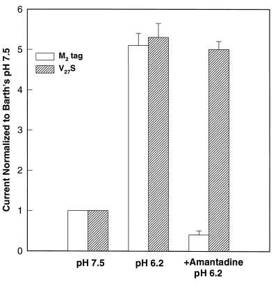
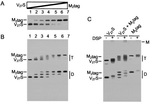
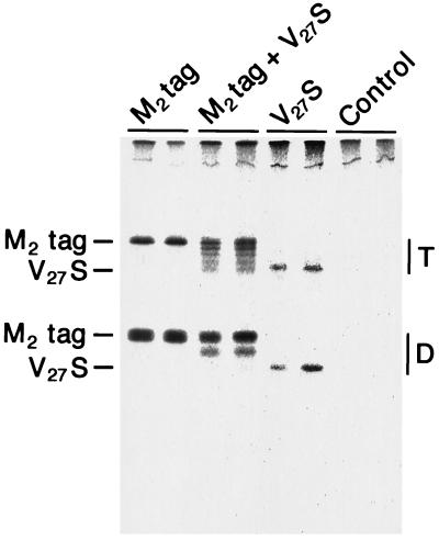
The influenza A virus M2 integral membrane protein is an ion channel that permits protons to enter virus particles during uncoating of virions in endosomes and also modulates the pH of the trans-Golgi network in virus-infected cells. The M2 protein is a homo-oligomer of 97 residues, and analysis by chemical cross-linking and SDS/PAGE indicates M2 forms a tetramer. However, a higher order molecular form is sometimes observed and, thus, it is necessary to determine the active form of the molecule. This was done by studying the currents of oocytes that expressed mixtures of the wild-type M2 protein (epitope tagged) and the mutant protein M2-V27S, which is resistant to the inhibitor amantadine. The composition of mixed oligomers of the two proteins expressed at the plasma membrane of individual oocytes was quantified after antibody capture of the cell surface expressed molecules and it was found that the subunits mixed freely. When the ratio of wild-type to mutant protein subunits was 0. 85:0.15, the amantadine sensitivity was reduced to 50% and for a ratio of 0.71:0.29 to 20%. These results are consistent with the amantadine-resistant mutant being dominant and the oligomeric state being a tetramer.
Figures
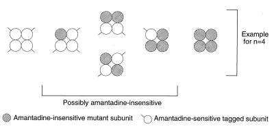
 ) assuming that the
active oligomer is a tetramer. All possible combinations of the two
subunits in a tetrameric channel are shown. As shown in Fig. 2, all
five species can be identified.
) assuming that the
active oligomer is a tetramer. All possible combinations of the two
subunits in a tetrameric channel are shown. As shown in Fig. 2, all
five species can be identified.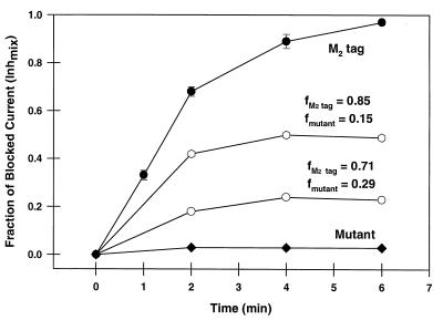
References
-
- Hay A J. Semin Virol. 1992;3:21–30.
-
- Lamb R A, Holsinger L J, Pinto L H. In: The Influenza A Virus M2 Ion Channel Protein and its Role in the Influenza Virus Life Cycle. Wimmer E, editor. Plainview, NY: Cold Spring Harbor Lab. Press; 1994. pp. 303–321.
-
- Pinto L H, Holsinger L J, Lamb R A. Cell. 1992;69:517–528. - PubMed
Publication types
MeSH terms
Substances
Grants and funding
LinkOut - more resources
Full Text Sources
Other Literature Sources