. 1997 May 13;94(10):5141-6.
doi: 10.1073/pnas.94.10.5141.

Ligand-dependent development of the endothelial and hemopoietic lineages from embryonic mesodermal cells expressing vascular endothelial growth factor receptor 2

Affiliations

Ligand-dependent development of the endothelial and hemopoietic lineages from embryonic mesodermal cells expressing vascular endothelial growth factor receptor 2

A Eichmann et al. Proc Natl Acad Sci U S A. .

Abstract

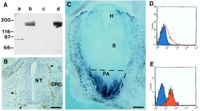
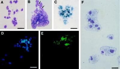
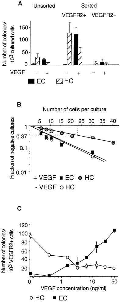
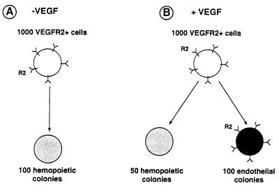
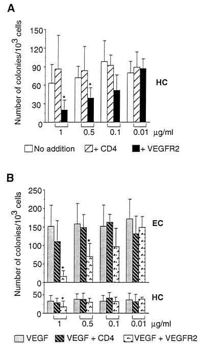
The existence of a common precursor for endothelial and hemopoietic cells, termed the hemangioblast, has been postulated since the beginning of the century. Recently, deletion of the endothelial-specific vascular endothelial growth factor receptor 2 (VEGFR2) by gene targeting has shown that both endothelial and hemopoietic cells are absent in homozygous null mice. This observation suggested that VEGFR2 could be expressed by the hemangioblast and essential for its further differentiation along both lineages. However, it was not possible to exclude the hypothesis that hemopoietic failure was a secondary effect resulting from the absence of an endothelial cell microenvironment. To distinguish between these two hypotheses, we have produced a mAb directed against the extracellular domain of avian VEGFR2 and isolated VEGFR2+ cells from the mesoderm of chicken embryos at the gastrulation stage. We have found that in clonal cultures, a VEGFR2+ cell gives rise to either a hemopoietic or an endothelial cell colony. The developmental decision appears to be regulated by the binding of two different VEGFR2 ligands. Thus, endothelial differentiation requires VEGF, whereas hemopoietic differentiation occurs in the absence of VEGF and is significantly reduced by soluble VEGFR2, showing that this process could be mediated by a second, yet unidentified, VEGFR2 ligand. These observations thus suggest strongly that in the absence of the VEGFR2 gene product, the precursors of both hemopoietic and vascular endothelial lineages cannot survive. These cells therefore might be the initial targets of the VEGFR2 null mutation.


Figures

Figure 1
Figure 1
Production of mAbs recognizing avian VEGFR2 and cell sorting of PA mesodermal cells. (A) Western blot with control CD4-Fc protein (10 μg, lanes a and c) and VEGFR2-Fc protein (10 μg, lanes b and d) was probed with anti-VEGFR2 (4H11) hybridoma supernatant (lanes c and d) and with anti-Fc mAb (lanes a and b). Anti-VEGFR2 recognizes only VEGFR2-Fc (lane d). (B) Immunohistochemistry with anti-VEGFR2 mAb on cryostat sections (20 μm) of an E4 chicken embryo. Section at the trunkal level showing anti-VEGFR2+ endothelial cells of the perineural vascular plexus (arrowheads). NT, neural tube; DRG, dorsal root ganglion. (Bar: 75 μm.) (C) Whole-mount in situ hybridization of a quail embryo at the 1-ss with a VEGFR2 antisense riboprobe. Note abundant positive cells in the mesoderm of the PA. H, headfold; S, somite (out of focus). The stippled region of the embryo was dissected. (Bar: 417 μm.) (D and E) Flow cytometry of VEGFR2+ cells from the PA. Fluorescence histogram showing anti-VEGFR2 labeling (red) before (D) and after (E) cell sorting.
Figure 2
Figure 2
Pheno types of hemopoietic cells obtained from VEGFR2+ PA cells. (A and B) MGG staining of hemopoietic colonies of thrombocyte (A) and thromboblast/erythroblast (B) type. Arrows in B point to two erythrocytes. (Bar: 25 μm.) (C) Benzidine-positive colony belonging to the erythrocytic lineage. (Bar: 21 μm.) (D) Hoechst nuclear stain and (E) CD 41/61 staining of a thromboblastic colony. (Bar: 26 μm.) (F) MGG staining of macrophages obtained in the presence of fibroblast-conditioned medium. (Bar: 18 μm.)
Figure 3
Figure 3
Phenotypes of colonies obtained in the presence of VEGF. (A) Typical appearance of a culture. MGG staining of two colonies, one hemopoietic (HC) of erythroblast/thromboblast phenotype, the other (EC) of endothelial phenotype. (Bar: 28 μm.) (BG) VEGF-induced colonies consist of endothelial cells. (B) Anti-VEGFR2 mAb staining followed by alkaline phosphatase-coupled secondary antibody. (Bar: 40 μm.) (C) In situ hybridization with a VEGFR3 antisense riboprobe. (Bar: 16 μm.) (D) Hoechst nuclear stain and (E) DiI-Ac-LDL uptake of an endothelial cell colony. (Bar: 32 μm.) (F) Hoechst nuclear stain and (G) mAb 118 immunoreactivity. (Bar: 53 μm.)
Figure 4
Figure 4
(A) Colony-forming cells (CFC) of the PA are enriched in the VEGFR2+ population as compared with unsorted or VEGFR2− cells. HC, hemopoietic colonies; EC, endothelial colonies. Each column represents the mean number of CFC calculated per 1 × 10³ cells obtained in three independent sorting experiments ± SD. (B) Limiting dilution analysis of VEGFR2+ cells. One of two experiments showing similar results. The frequency of colony-initiating progenitors at 37% negative wells was, according to the Poisson analysis, 1/22 for HC and 1/10 for EC in the presence of VEGF and 1/11 for HC obtained without VEGF. (C) Dose-dependent stimulation of endothelial cell development by VEGF. Values represent the mean ± SD from two experiments.
Figure 5
Figure 5
Schematic representation of the results. One thousand VEGFR2+ cells cultured in the absence (A) or presence (B) of VEGF give rise to endothelial and hemopoietic colonies as indicated. Endothelial colonies only maintain VEGFR2 expression.
Figure 6
Figure 6
(A) Soluble VEGFR2 inhibits hemopoietic colony formation. One thousand VEGFR2+ cells were cultured without growth factor addition and in the presence of the indicated concentrations of soluble VEGFR2-Fc protein or CD4-Fc protein. Values represent the mean ± SD from three experiments. Statistical analysis was performed using the paired Student’s t test. ∗ indicates P ≤ 0.05. (B) Soluble VEGFR2 inhibits VEGF-induced colony formation. One thousand VEGFR2+ cells were cultured in the presence of 50 ng/ml VEGF and the indicated concentrations of soluble VEGFR2-Fc protein or CD4-Fc protein. Values represent the mean ± SD from four (1 μg/ml and 0.1 μg/ml) or three experiments. Statistical analysis was performed as in A. HC, hemopoietic colonies, EC, endothelial colonies.

References

    1. Sabin F R. Contributions to Embryology. Vol. 9. Carnegie Institution; 1920. , No. 36, pp. 214–262.
    1. Murray P D F. Proc R Soc (London) Ser B. 1932;11:497–521.
    1. Mustonen T, Alitalo K. J Cell Biol. 1995;129:895–898. - PMC - PubMed
    1. Shibuya M. Adv Cancer Res. 1995;67:281–316. - PubMed
    1. Fong G H, Rossant J, Gertsenstein M, Breitman M L. Nature (London) 1995;376:66–70. - PubMed

Publication types

MeSH terms

Substances