. 1997 May 13;94(10):5162-6.
doi: 10.1073/pnas.94.10.5162.

The origin and evolution of animal appendages

Affiliations

The origin and evolution of animal appendages

G Panganiban et al. Proc Natl Acad Sci U S A. .

Abstract

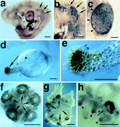
Animals have evolved diverse appendages adapted for locomotion, feeding and other functions. The genetics underlying appendage formation are best understood in insects and vertebrates. The expression of the Distal-less (Dll) homeoprotein during arthropod limb outgrowth and of Dll orthologs (Dlx) in fish fin and tetrapod limb buds led us to examine whether expression of this regulatory gene may be a general feature of appendage formation in protostomes and deuterostomes. We find that Dll is expressed along the proximodistal axis of developing polychaete annelid parapodia, onychophoran lobopodia, ascidian ampullae, and even echinoderm tube feet. Dll/Dlx expression in such diverse appendages in these six coelomate phyla could be convergent, but this would have required the independent co-option of Dll/Dlx several times in evolution. It appears more likely that ectodermal Dll/Dlx expression along proximodistal axes originated once in a common ancestor and has been used subsequently to pattern body wall outgrowths in a variety of organisms. We suggest that this pre-Cambrian ancestor of most protostomes and the deuterostomes possessed elements of the genetic machinery for and may have even borne appendages.


Figures

Figure 1
Figure 1
Dll expression in representative protostomes. (a) Lateral view of a late stage Precis coenia butterfly embryo stained with the Dll antibody. Arrows (➞) point to the distal tips of the left abdominal prolegs. Dll expression is detected in central nervous system in the brain (br) and in the ventral nerve cord (➤). (b) Higher magnification image of abdominal prolegs (➞) from an embryo similar to that shown in a. (c) Ventral view of a late stage Peripatopsis capensis onychophoran embryo stained with the Dll antibody. The antennae (ant), oral papilla (➤) and lobopods express Dll. The lobopods shown in higher magnification in e are indicated with ➞. (d) Right halves of two segments from a young P. capensis embryo stained with the Dll antibody. Dll expression is detected in the ectoderm of the presumptive lobopods prior to the formation of visible buds. (e) Higher magnification view of the lobopods indicated in d. (∗) The neurogenic ectoderm, which also expresses Dll. (f) Polychaete annelid Chaetopterus variopedatus, ventral view of larva just prior to metamorphosis. Dll expressing cells are visible in parapodial rudiments (➞), antennae (out of focus on opposite dorsal surface, (➤), prospective feeding organs (⊓), and in the neurogenic ectoderm (∗). (g) C.variopedatus, same specimen at a higher magnification, showing Dll reactive ectodermal nuclei in prospective distal cells of the anterior parapodia (➞) and in the neurogenic region (∗). (h) Later stage C. variopedatus larva showing staining in distal portions of two anterior parapodia (➞). Anterior is to the left in all panels. (Bars = 0.1 mm.)
Figure 2
Figure 2
Dll expression in representative deuterostomes. (a) Nine-day mouse embryo stained with the Dll antibody. Arrows (➞) point to medial border of cells expressing one or more Dlx genes in the presumptive forelimb. Dlx expression can be detected in developing mouse limbs as the bud forms from the flank, and somewhat earlier than previously reported for mice or other vertebrates (–11). (b) Higher magnification view of the forelimb indicated in A. (c) Dorsal view of the forelimb of a 10-day mouse embryo stained with the Dll antibody. (➤) The position of the apical ectodermal ridge. (d) Three-day Molgula occidentalis ascidian larva from which an ampulla is extending. Cells at the distal tip of the ampulla express Dll (➞). (e) Higher magnification view of the ampulla shown in d. (f and g) Metamorphosing Strongylocentrotus droebachiensis sea urchin larvae stained with Dll antibody. Cells at the distal tip of the tube feet (➞) express Dll prior to (f) and during (g) extension from the body wall. (h) Higher magnification view of a tube foot (➞) and spines (➤) from an S. droebachiensis larva similar to that shown in g. Cells at the distal tip of the developing spines, as well as the tube feet express Dll. (Bars = 0.1 mm.)
Figure 3
Figure 3
The evolution of animal appendages. The cladogram depicts the relationships between a selected subset of animal phyla and is based upon a combination of sources (see ref. for review; other phylogenies are possible but do not alter the basic inferences drawn here). Branch lengths are not scaled. Taxa for which Dll expression data is presented here are shown in bold along with the appearance of the various appendages in these groups. The appearance of various appendages within the phyla studied here are indicated. Only the jointed limbs of arthropods and tetrapod limbs arose by modification of a pre-existing appendage. The immediate ancestors of polychaetes, Onychophora, echinoderms, urochordates, and vertebrates are not thought to have borne appendages that would be homologous to the structures analyzed here. The deuterostome ancestor (b) may have had one Dll/Dlx gene since cephalochordates have only one (13). However, the presence of two Dlx genes in ascidians (15) leaves open the possibility that at least one Dlx duplication event occurred earlier in the deuterostome lineage. The ancestor of the appendage-bearing protostome clade and the deuterostomes possessed the Dll gene and all of the features of the proposed Urbilaterian (18), and may have borne appendages (a). The Dll gene predates this ancestor and is found in nematodes and expressed in the CNS, which may be an older site of function than body wall outgrowths. There is a report of a possible cnidarian Dll ortholog (19), but the similarity of the cnox3 homeodomain to Drosophila Dll (56%) is not significantly higher than that of nonorthologous homeodomains, and the Dll antibody does not recognize this gene product. We therefore believe the origin of Dll is unresolved.
Figure 4
Figure 4
Identification and expression of a nematode Dll gene. (a) Alignment of the homeodomains of Drosophila (52), mouse (48, 53), and nematode Dll/Dlx (50) gene products. The amino acid sequence of the Ce-Dll homeodomain is 74% identical to that of Drosophila Dll. (➤), The position of an intron conserved between the fly (54), ascidian (15), and nematode (50) genes. (b and c) Lateral views of 100-cell (b) and comma (c) stage Caenorhabditis elegans embryos stained with the Dll antibody (green) and an antibody (O1C1D4) that recognizes P-granules (blue). (d) Dorsolateral view of a three-fold stage C. elegans embryo stained with the Dll antibody (green) and an antibody (3NB12) that recognizes pharyngeal cells (red). The Ce-Dll expressing cells in b and c are the precursors of the nerve ring cells that express Ce-Dll in d. Anterior is to the left in b–d, and the magnification in b–d is the same. (Bar = 10 μm.)

References

    1. Wang B B, Muller-Immergluck M M, Austin J, Robinson N T, Chisholm A, Kenyon C. Cell. 1993;74:29–42. - PubMed
    1. Cohen S M. Nature (London) 1990;343:173–177. - PubMed
    1. Panganiban G, Nagy L, Carroll S B. Curr Biol. 1994;4:671–675. - PubMed
    1. Panganiban G, Sebring A, Nagy L, Carroll S B. Science. 1995;270:1363–1366. - PubMed
    1. Popadic A, Rusch D, Peterson M, Rogers B T, Kaufman T C. Nature (London) 1996;380:395.

Publication types

MeSH terms

Substances

LinkOut - more resources