. 1997 May 13;94(10):5207-12.
doi: 10.1073/pnas.94.10.5207.

Functional domains are specified to single-cell resolution in a Drosophila epithelium

Affiliations

Functional domains are specified to single-cell resolution in a Drosophila epithelium

M A Sözen et al. Proc Natl Acad Sci U S A. .

Abstract

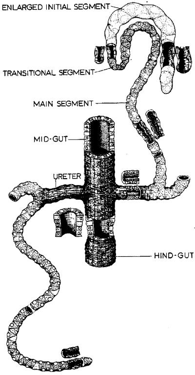
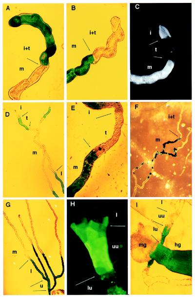
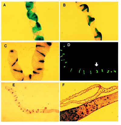
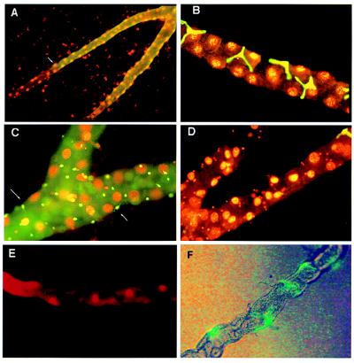
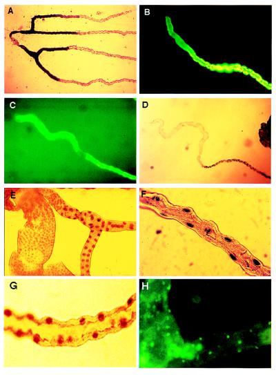
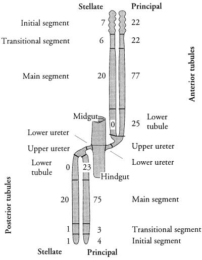
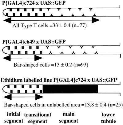
Specification of pattern is fundamental to the development of a multicellular organism. The Malpighian (renal) tubule of Drosophila melanogaster is a simple epithelium that proliferates under the direction of a single tip cell into three morphologically distinct domains. However, systematic analysis of a panel of over 700 P[GAL4] enhancer trap lines reveals unexpected richness for such an apparently simple tissue. Using numerical analysis, it was possible formally to reconcile apparently similar or complementary expression domains and thus to define at least five genetically defined domains and multiple cell types. Remarkably, the positions of domain boundaries and the numbers of both principal and secondary ("stellate") cell types within each domain are reproducible to near single-cell precision between individual animals. Domains of physiological function were also mapped using transport or expression assays. Invariably, they respect the boundaries defined by enhancer activity. These genetic domains can also be visualized in vivo, both in transgenic and wild-type flies, providing an "identified cell" system for epithelial physiology. Building upon recent advances in Drosophila Malpighian tubule physiology, the present study confirms this tissue as a singular model for integrative physiology.


Figures

Figure 1
Figure 1
Classical morphology of the D. melanogaster Malpighian tubule. [Reproduced with permission from ref. (Copyright 1978, Academic Press).]
Figure 2
Figure 2
Genetic mapping of tubule subregions. (A) Line 155Y marks the initial and transitional segments of posterior tubules by inclusion, whereas (B) lines c709 and c776 mark them by exclusion. Line c825 shows staining of the initial and main, but not transitional, segments in posterior (C) and anterior (E) tubules. (D) In contrast, line c507 marks the transitional segment and lower tubule, but not the initial or main segments (anterior tubule). (F) Line c374 marks the main segment. In this line, the boundary with the transitional segment is clear, but the transition to lower tubule is gradual (see Fig. 3). (G) Line c507 marks the lower tubule and ureter. (H) Line c649 marks an “upper ureter” domain from the tubule bifurcation to halfway along the ureter (anti-β-galactosidase fluorescence). (I) Line c601 marks the complementary lower ureter and hindgut. The diameter of the tubule can be taken as 35 μm throughout. i, Initial segment; t, transitional segment; m, main segment; l, lower tubule; u, ureter; uu, upper ureter; lu, lower ureter; mg, midgut; hg, hindgut.
Figure 3
Figure 3
Genetic mapping of tubule cell types. (A) Lines c324 and c374 distinguish subsets of morphologically indistinguishable principal epithelial cells in the main segment. (B) Lines c710 and c724 are expressed uniquely in the stellate cells of adults and larvae. (C) Lines c710 and c724 also mark bar-shaped cells in the initial segment of anterior tubules. (D) Type II cell morphology switches from bar-shaped to stellate at the transitional–main tubule boundary (arrow). The tip of the tubule is to the left. (E) Line c649 marks only the bar-shaped type II cells of anterior tubule initial and transitional segments. (F) Line c325 demonstrates the presence of a small, previously unreported cell type in the lower tubules and ureter and of homologues in the posterior midgut.
Figure 4
Figure 4
Counterstaining with ethidium bromide allows cell numbers to be counted. (A) Reporter gene expression was detected with fluorescein-coupled anti-β-galactosidase secondary antibody (green) in line c507 and counterstained with ethidium bromide. The boundary of β-galactosidase expression can be identified to single-cell resolution, and the number of principal cell nuclei to the tubule bifurcation can be counted. (B) Line c724 shows the relative abundance of principal and stellate cells in the main segment. In addition, the nuclei of the stellate cells are clearly smaller than those of principal cells. (C) Line c710 shows staining of the tiny cells of the ureter. Fine processes are visible in some cells (arrows). (D) Line c724, showing that stellate cells do not overlap with tiny cells in the lower tubule. A single stained stellate cell is visible at the junction of the lower tubule, and the tiny cell nuclei are visible in the lower tubule (cf. C). (E) Vital staining of tubule main segment principal cell nuclei with 10 μg/ml ethidium bromide for 5 min and viewed by epifluorescence. (F) Vital labeling of stellate cells with genetically modified (S65T) GFP. Flies carrying the P{UAS-GFP.S65T}T10 construct (Bloomington stock) were crossed to P{GAL4} line c724, and red-eyed adults were dissected in Drosophila saline and viewed without further treatment with mixed bright-field and fluorescein optics.
Figure 5
Figure 5
Functional mapping of tubule regions. (A) Alkaline phosphatase staining of lower tubule. Tubules were fixed and stained for alkaline phosphatase. This shows a perfect overlap with line c507 (cf. Fig. 2G). (B and C) Short-term (10 min) labeling with 10 μg/ml rhodamine 123 identifies the main segment of both posterior (B) and anterior (C) tubules as the region responsible for transport of this organic molecule. (D) Initial and transitional segments express V-ATPase at a far lower level than the main segment, as detected by a reporter gene inserted into the vha55 gene in the P-element lethal line vha55j2e5 (28). This corresponds to the boundary reported by lines 155Y, c709, and c776 in Fig. 2 A and B. (E) A boundary midway along the ureter is marked by a change in the size of nuclei labeled in line vha55j2e5. This corresponds to the boundary reported by lines c649 and c601 in Fig. 2 H and I. (F) Within the main segment, only principal cell nuclei (cf. Fig. 4B) are labeled in line vha55j2e5. (G) Within the main segment, only principal cell nuclei (cf. Fig. 4B) are labeled in P-element line l(2)k02508, a recessive lethal insertion within the gene encoding the body-enriched vha68–2 V-ATPase A subunit (36). (H) The tiny cells (cf. Figs. 3F and 4C) are labeled by antibodies to horseradish peroxidase, which recognize an epitope on a Drosophila nervous system-specific Na+,K+ ATPase β-subunit (37).
Figure 6
Figure 6
Summary of tubule regional architecture. Regions defined in this study are labeled (cf. Fig. 1); the numbers of principal and stellate cells in each region are shown. Numerical data are derived from Table 2; SEs are not shown, as they are <1 in each case.
Figure 7
Figure 7
Numerical reconciliation of genetic and functional domains. (Top) Line c724 drives GFP expression in 33 type II cells of initial, transitional, and main segments. (Middle) Line c649 drives expression in 13 bar-shaped cells, diagnostic of initial and transitional segments (Table 2). (Bottom) When ethidium bromide is added to c724, it is transported into the cells, obscuring the view of all but the most distal 13 cells. This shows that the organic cation transport system operates in a compartment statistically indistinguishable from main segment.

References

    1. Bertram M J, Akerkar G A, Ard R L, Gonzalez C, Wolfner M F. Mech Dev. 1992;38:33–40. - PubMed
    1. Hartenstein V, Jan Y N. Roux’s Arch Dev Biol. 1992;201:194–220. - PubMed
    1. Riesgo-Escovar J, Woodard C, Gaines P, Carlson J. J Neurobiol. 1992;23:947–964. - PubMed
    1. Doe C Q, ChuLaGraff Q, Wright D M, Scott M P. Cell. 1991;65:451–464. - PubMed
    1. Nose A, Mahajan V B, Goodman C S. Cell. 1992;70:553–567. - PubMed

Publication types

MeSH terms

LinkOut - more resources