. 1997 May 13;94(10):5261-6.
doi: 10.1073/pnas.94.10.5261.

A novel pair of immunoglobulin-like receptors expressed by B cells and myeloid cells

Affiliations

A novel pair of immunoglobulin-like receptors expressed by B cells and myeloid cells

H Kubagawa et al. Proc Natl Acad Sci U S A. .

Abstract

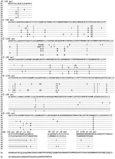
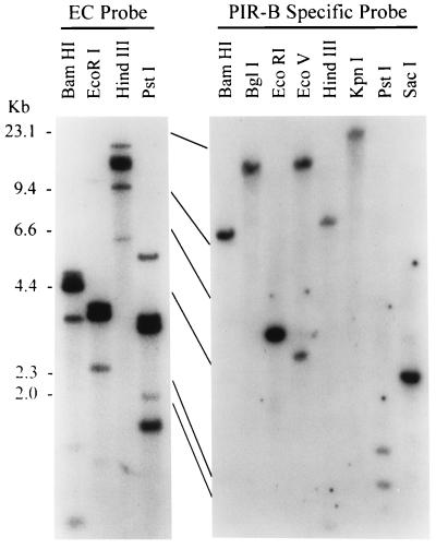
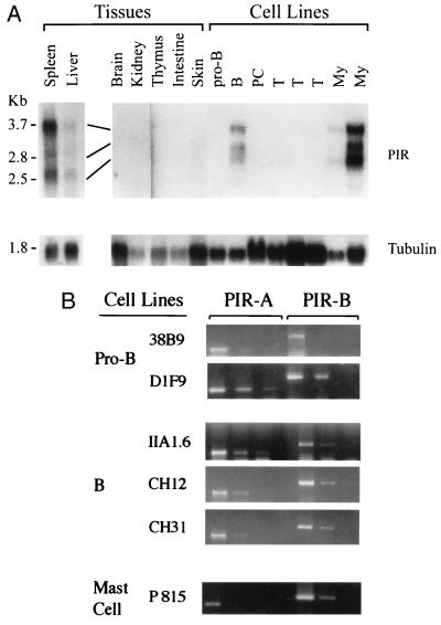
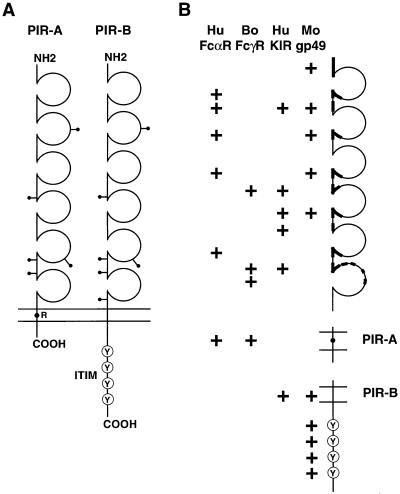
An Fcalpha receptor probe of human origin was used to identify novel members of the Ig gene superfamily in mice. Paired Ig-like receptors, named PIR-A and PIR-B, are predicted from sequence analysis of the cDNAs isolated from a mouse splenic library. Both type I transmembrane proteins possess similar ectodomains with six Ig-like loops, but have different transmembrane and cytoplasmic regions. The predicted PIR-A protein has a short cytoplasmic tail and a charged Arg residue in the transmembrane region that, by analogy with the FcalphaR relative, suggests the potential for association with an additional transmembrane protein to form a signal transducing unit. In contrast, the PIR-B protein has an uncharged transmembrane region and a long cytoplasmic tail containing four potential immunoreceptor tyrosine-based inhibitory motifs. These features are shared by the related killer inhibitory receptors. PIR-A proteins appear to be highly variable, in that predicted peptide sequences differ for seven randomly selected PIR-A clones, whereas PIR-B cDNA clones are invariant. Southern blot analysis with PIR-B and PIR-A-specific probes suggests only one PIR-B gene and multiple PIR-A genes. The PIR-A and PIR-B genes are expressed in B lymphocytes and myeloid lineage cells, wherein both are expressed simultaneously. The characteristics of the highly-conserved PIR-A and PIR-B genes and their coordinate cellular expression suggest a potential regulatory role in humoral, inflammatory, and allergic responses.


Figures

Figure 1
Figure 1
Sequence diversity among members of the activation-type (PIR-A) receptors. Variable amino acid sequences (one-letter code) deduced from the seven randomly picked, PIR activation-type cDNA clones (A1 to A7) are aligned with the invariable peptide sequence of the PIR inhibitory-type cDNA clone (B1). Amino acid identity is indicated by dashes (–); a change, by boldface letters; and a deletion introduced for optimal alignment, by slashes (/). A charged Arg residue in the transmembrane region of PIR-A-type clones and the ITIM-like motifs in the cytoplasmic tail of PIR-B clone are also in boldface letters. Sequences are divided into each putative region: SP, signal peptide; EC, extracellular domain; ECmp, membrane proximal extracellular; TM, transmembrane; and CY, cytoplasmic regions. The boundaries of EC1/EC2 to EC5/EC6 were established from the analysis of genomic clones. Numbers in parentheses indicate the amino acid (aa) length of each region.
Figure 2
Figure 2
Southern blot analysis of PIR gene family. DNA from BALB/c testis was analyzed with PIR probes corresponding to the common extracellular (EC) region (Left) or to the PIR-B-specific region (Right).
Figure 3
Figure 3
Chromosomal localization of the PIR gene family. Partial chromosome 7 linkage map showing the location of Pir in relation to linked genes. cM, centiMorgans.
Figure 4
Figure 4
Tissue specificity and cell lineage restriction of PIR gene expression. (A) RNA blot analysis. One microgram of poly(A)+ RNA (spleen, liver) or 10 μg of total RNA from the indicated murine tissues and cell lines was analyzed with 32P-labeled PIR (Upper) and tubulin (Lower) probes. Similar transcripts were also observed in bone marrow. (B) RT-PCR analysis. Three different concentrations of first-strand cDNA products were subjected to PCR amplification of PIR-A, PIR-B, and actin transcripts (not shown). Amplified products electrophoresed in 2% agarose were stained with ethidium bromide. Similar coexpression patterns were observed for macrophage cell lines.
Figure 5
Figure 5
Model of PIR-A and PIR-B and their relationship with other Ig gene superfamily members. (A) PIR-A and PIR-B cDNA clones encode type I transmembrane proteins with similar extracellular domains, but different transmembrane and cytoplasmic regions. The extracellular region has six Ig-like domains, the second of which is V-like and the others C2-like. There are five or six potential sites for N-linked glycosylation (—•). The predicted PIR-A protein contains a relatively short cytoplasmic tail and a charged arginine (R) residue in the transmembrane region, whereas the PIR-B protein has a typical nonpolar transmembrane region and a long cytoplasmic tail with four potential tandem immunoreceptor tyrosine-based inhibitory motifs (Y). (B) Schematic depiction of the regions of PIR-A and PIR-B that share significant homology with other members of the Ig gene superfamily, the human FcαR (8), bovine FcγR (42), human KIRs (–41), and mouse gp49 (43).

Comment in

  • Inhibitory receptors abound?
    Cambier JC. Cambier JC. Proc Natl Acad Sci U S A. 1997 Jun 10;94(12):5993-5. doi: 10.1073/pnas.94.12.5993. Proc Natl Acad Sci U S A. 1997. PMID: 9177155 Free PMC article. Review. No abstract available.

References

    1. Ravetch J V. Cell. 1994;78:553–560. - PubMed
    1. Jouvin M-H, Numerof R P, Kinet J-P. Semin Immunol. 1995;7:29–35. - PubMed
    1. Weiss A, Littman D R. Cell. 1994;76:263–274. - PubMed
    1. Cambier J C. J Immunol. 1995;155:3281–3285. - PubMed
    1. Albrechtsen M, Yeaman G R, Kerr M A. Immunology. 1988;64:201–206. - PMC - PubMed

Publication types

MeSH terms