

Apoptosis within spontaneously accepted mouse liver allografts: evidence for deletion of cytotoxic T cells and implications for tolerance induction
- PMID: 9144477
- PMCID: PMC2954768
Apoptosis within spontaneously accepted mouse liver allografts: evidence for deletion of cytotoxic T cells and implications for tolerance induction
Abstract
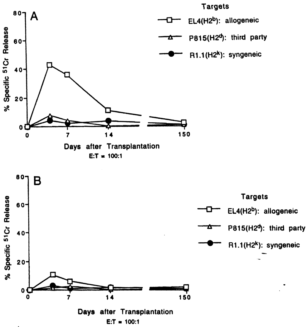
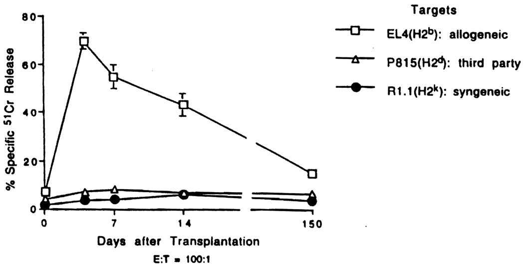
MHC-mismatched liver grafts are accepted spontaneously between many mouse strains. The underlying mechanism(s) is unclear. In the B10 (H2(b)) to C3H (H2(k)) strain combination used in this study, donor T cells within the liver were rapidly replaced within 2 to 4 days of transplantation with those of the recipient. Freshly isolated liver graft-infiltrating cells harvested on days 4 and 7 exhibited strong CTL responses against donor alloantigens. CTL activity was reduced substantially, however, by day 14, although levels of CTL precursors in the spleen and liver remained high. Examination of the liver allografts by in situ terminal deoxynucleotidyltransferase-catalyzed dUTP-digoxigenin nick end labeling on days 4, 7, and 14 after transplantation revealed prominent apoptotic cells dispersed throughout the nonparenchymal cell population. When acute liver allograft rejection was induced by administration of IL-2 from days 0 to 4 post-transplant (median survival time, 5 days), apoptotic activity (day 4) was reduced substantially, whereas CTL activity was enhanced. Nonparenchymal cells isolated from allografts of unmodified recipients 4, 7, and 14 days after transplantation exhibited significantly higher DNA fragmentation after 18-h culture than cells from liver isografts. Moreover, the level was 4 to 5 times higher than that of cells from IL-2-treated mice (on day 4). These observations suggest that T cell deletion, not regulation, may be responsible for spontaneous liver allograft acceptance. The molecular recognition events that cause apoptosis of infiltrating T cells and why this occurs within liver grafts, but not heart or skin grafts, remain to be elucidated.
Figures


Similar articles
-
Increased apoptosis of immunoreactive host cells and augmented donor leukocyte chimerism, not sustained inhibition of B7 molecule expression are associated with prolonged cardiac allograft survival in mice preconditioned with immature donor dendritic cells plus anti-CD40L mAb.Transplantation. 1999 Sep 27;68(6):747-57. doi: 10.1097/00007890-199909270-00006. Transplantation. 1999. PMID: 10515374 Free PMC article.
-
Il-12 antagonism enhances apoptotic death of T cells within hepatic allografts from Flt3 ligand-treated donors and promotes graft acceptance.J Immunol. 2001 May 1;166(9):5619-28. doi: 10.4049/jimmunol.166.9.5619. J Immunol. 2001. PMID: 11313402
-
Donor pretreatment with Flt-3 ligand augments antidonor cytotoxic T lymphocyte, natural killer, and lymphokine-activated killer cell activities within liver allografts and alters the pattern of intragraft apoptotic activity.Transplantation. 1998 Jun 27;65(12):1590-8. doi: 10.1097/00007890-199806270-00009. Transplantation. 1998. PMID: 9665075 Free PMC article.
-
Prevention and restoration of second-set liver allograft rejection in presensitized mice: the role of "passenger" leukocytes, donor major histocompatibility complex antigens, and host cytotoxic effector mechanisms.Transplantation. 1999 Feb 15;67(3):444-50. doi: 10.1097/00007890-199902150-00018. Transplantation. 1999. PMID: 10030293
-
Costimulation blockade promotes the apoptotic death of graft-infiltrating T cells and prolongs survival of hepatic allografts from FLT3L-treated donors.Transplantation. 2001 Oct 27;72(8):1423-32. doi: 10.1097/00007890-200110270-00016. Transplantation. 2001. PMID: 11685115
Cited by
-
Antithymocyte globulin facilitates alloreactive T-cell apoptosis by means of caspase-3: potential implications for monitoring rejection-free outcomes.Transplantation. 2015 Jan;99(1):164-70. doi: 10.1097/TP.0000000000000289. Transplantation. 2015. PMID: 25531894 Free PMC article.
-
Increased apoptosis of immunoreactive host cells and augmented donor leukocyte chimerism, not sustained inhibition of B7 molecule expression are associated with prolonged cardiac allograft survival in mice preconditioned with immature donor dendritic cells plus anti-CD40L mAb.Transplantation. 1999 Sep 27;68(6):747-57. doi: 10.1097/00007890-199909270-00006. Transplantation. 1999. PMID: 10515374 Free PMC article.
-
Immunomodulatory Role of Kupffer Cell in Liver Allografts.Comp Hepatol. 2004 Jan 14;3 Suppl 1(Suppl 1):S32. doi: 10.1186/1476-5926-2-S1-S32. Comp Hepatol. 2004. PMID: 14960184 Free PMC article. No abstract available.
-
Single-Cell RNA Sequencing of Donor-Reactive T Cells Reveals Role of Apoptosis in Donor-Specific Hyporesponsiveness of Kidney Transplant Recipients.Int J Mol Sci. 2023 Sep 23;24(19):14463. doi: 10.3390/ijms241914463. Int J Mol Sci. 2023. PMID: 37833911 Free PMC article.
-
Graft-infiltrating PD-L1hi cross-dressed dendritic cells regulate antidonor T cell responses in mouse liver transplant tolerance.Hepatology. 2018 Apr;67(4):1499-1515. doi: 10.1002/hep.29529. Epub 2018 Feb 18. Hepatology. 2018. PMID: 28921638 Free PMC article.
References
Publication types
MeSH terms
Substances
Grants and funding
LinkOut - more resources
Full Text Sources
Medical
Research Materials