. 1997 May 5;137(3):539-53.
doi: 10.1083/jcb.137.3.539.

A proteasome cap subunit required for spindle pole body duplication in yeast

Affiliations

A proteasome cap subunit required for spindle pole body duplication in yeast

H B McDonald et al. J Cell Biol. .

Abstract

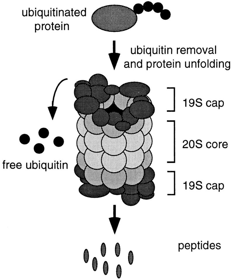
Proteasome-mediated protein degradation is a key regulatory mechanism in a diversity of complex processes, including the control of cell cycle progression. The selection of substrates for degradation clearly depends on the specificity of ubiquitination mechanisms, but further regulation may occur within the proteasomal 19S cap complexes, which attach to the ends of the 20S proteolytic core and are thought to control entry of substrates into the core. We have characterized a gene from Saccharomyces cerevisiae that displays extensive sequence similarity to members of a family of ATPases that are components of the 19S complex, including human subunit p42 and S. cerevisiae SUG1/CIM3 and CIM5 products. This gene, termed PCS1 (for proteasomal cap subunit), is identical to the recently described SUG2 gene (Russell, S.J., U.G. Sathyanarayana, and S.A. Johnston. 1996. J. Biol. Chem. 271:32810-32817). We have shown that PCS1 function is essential for viability. A temperature-sensitive pcs1 strain arrests principally in the second cycle after transfer to the restrictive temperature, blocking as large-budded cells with a G2 content of unsegregated DNA. EM reveals that each arrested pcs1 cell has failed to duplicate its spindle pole body (SPB), which becomes enlarged as in other monopolar mutants. Additionally, we have shown localization of a functional Pcs1-green fluorescent protein fusion to the nucleus throughout the cell cycle. We hypothesize that Pcs1p plays a role in the degradation of certain potentially nuclear component(s) in a manner that specifically is required for SPB duplication.


Figures

Figure 1
Figure 1
Schematic representation of a 26S proteasome. The 20S proteolytic barrel structure is indicated in pale grey and the 19S cap structures at either end are in dark grey. The caps are believed to recognize ubiquitinated proteins, unfold them, and feed them into the proteolytic core in an ATP-dependent manner, resulting in the release of peptides and free ubiquitin.
Figure 2
Figure 2
Organization and sequence analysis of the PCS1 gene. (A) Partial restriction map of the genomic region surrounding PCS1. The PCS1 and CDC31 coding regions are indicated by arrows beneath the map; ORF indicates a third open reading frame. The line above the map represents the sequenced region shown in B; this region is sufficient to supply PCS1 function in cells lacking other functional copies of the gene. H, HindIII; B, BamHI; R, EcoRI. (B) Nucleotide and predicted amino acid sequence of PCS1. A potential nuclear localization signal is boxed; a heptad repeat characteristic of α-helical coiledcoils is indicated by triangles; sequence motifs characteristic of an ATP-binding domain are underlined; and the site of gene disruption described in the text is indicated by an arrow under the first ATP motif. These sequence data are available from GenBank/EMBL/DDBJ under accession number U93262. The PCS1 sequence is also entered in these databases as SUG2 (accession number SCU43720 [GB]) (83) and in the S. cerevisiae genome database by J.H. McCusker (Duke University Medical Center, Durham, NC) and J.E. Haber (Brandeis University, Waltham, MA) as representing CRL13, which was originally identified by a temperature-sensitive, cycloheximide-resistant allele (65). (C) Structural analysis of Pcs1p. The top line is a graphical representation of the relative hydrophilicity of its amino acid sequence (57). The next three lines represent secondary structural predictions calculated by the method of Garnier et al. (29) and displayed in graphical form. Regions predicted to form turns, α helices, or β sheets are indicated by elevation of the appropriate line.
Figure 3
Figure 3
Multiple sequence alignment of Pcs1p, p42, Sug1/Cim3 protein, and S4. (Black boxes) Amino acid identity; (grey boxes) similarity. This display was created using the program BOXSHADE.
Figure 4
Figure 4
Characterization of pcs1td. (A) Growth at different temperatures of yeast strains containing PCS1 or pcs1td. Each panel represents duplicate patches of the following yeast strains: (a) Wx257-5c (PCS1), (b) YHM11.2 (pcs1td), (c) YHM13.1 (PCS1, ubr1Δ:: HIS3), and (d) YHM12.1 (pcs1td, ubr1Δ::HIS3). (B) Percentages of unbudded, small-budded, and large-budded cells in asynchronous PCS1 and pcs1td strains before and after shift to 37°C. (C and D) Percentages of cell types in synchronous PCS1 and pcs1td strains. A shift from 23° to 37°C occurred at time 0.
Figure 5
Figure 5
Flow cytometric analysis of pcs1td. (A) Wx257-5c (PCS1, open curve) compared with YHM11.2 (pcs1td, shaded curve) at 23°C. (B) PCS1 (open curve) compared with pcs1td (shaded curve) at 37°C. The left peak represents G1 cells and the right peak represents G2/M cells. In these histograms, the x-axis indicates relative DNA content measured by propidium iodide fluorescence, and the y-axis indicates the relative number of cells. Each sample represents 15,000 cells.
Figure 6
Figure 6
Cytological analysis of pcs1td cells using immunofluorescence microscopy. (Left) DNA staining (DAPI); (right) microtubule staining (FITC). (A) Wx257-5c (PCS1) at 37°C. (B–E) YHM11.2 (pcs1td) at 37°C. Intranuclear spindles appear to be absent in the mutant cells. Bar, 5 μm.
Figure 7
Figure 7
DNA localization in linked large-budded pcs1td cells at 37°C. (A) Nomarski image of cells. The numbers designate the presumed order of budding, as explained in the text. (B) DNA (DAPI). (C) Microtubules (FITC). Bar, 5 μm.
Figure 8
Figure 8
Spindle morphology in (A) YHM11.2 (pcs1td), (B) CMY763 (cim3-1), and (C) CMY765 (cim5-1) strains at 37°C. (Bars) Percentages of large-budded cells with the indicated spindle forms.
Figure 9
Figure 9
Electron micrographs of the YHM11.2 (pcs1td) mutant after transfer to 37°C. A normal mitotic spindle was seen in 16% of the cells analyzed, as shown in A. In this cell, the other SPB was located in a different section (not shown). The arrow indicates the halfbridge structure adjacent to the SPB. Pairs of images (B, B′ and C, C′) represent adjacent serial sections through the poles of monopolar spindles, as were seen in 84% of arrested cells. Bar, 0.5 μm.
Figure 10
Figure 10
GFP–Pcs1p expression and localization. (A) Western blot analysis of whole-cell extracts probed with anti-GFP antiserum. (Lane 1) Wx257-5c (PCS1). (Lane 2) YHM10.1.54 (GFP– PCS1). The presumptive GFP–Pcs1p fusion is indicated by the arrow, and size markers are in kD. (B and C) Fluorescence microscopy of YHM10.1.54 cells. (B) GFP (FITC channel). (1) Unbudded cell; (2) small-budded cell; (3 and 4) large-budded cells. (Asterisk) Less typical (∼5% of cells) appearance of GFP signal that is not coincident with nuclear DNA staining. (C) DNA (DAPI). Bar, 5 μm.
Figure 11
Figure 11
Sen3 protein localization. (A) MHY851 cells were stained with anti-HA antibody to visualize the Sen3 protein (FITC). (B) DNA (DAPI). Bar, 5 μm.

References

    1. Alber T. Structure of the leucine zipper. Curr Opin Gen Dev. 1992;2:205–210. - PubMed
    1. Altschul SF, Gish W, Miller W, Myers EW, Lipman DJ. Basic local alignment search tool. J Mol Biol. 1990;215:403–410. - PubMed
    1. Bachmair A, Finley D, Varshavsky A. In vivo half-life of a protein is a function of its amino-terminal residue. Science (Wash DC) 1986;234:179–186. - PubMed
    1. Baum P, Furlong C, Byers B. Yeast gene required for spindle pole body duplication: homology of its product with Ca2+-binding proteins. Proc Natl Acad Sci USA. 1986;83:5512–5516. - PMC - PubMed
    1. Boeke JD, LaCroute F, Fink GR. A positive selection for mutants lacking orotidine-5′-phosphate decarboxylase activity in yeast: 5-fluoro-orotic acid resistance. Mol Gen Genet. 1984;197:345–346. - PubMed

Publication types

MeSH terms