. 1997 May 5;137(3):595-608.
doi: 10.1083/jcb.137.3.595.

Differential sorting of lysosomal enzymes out of the regulated secretory pathway in pancreatic beta-cells

Affiliations

Differential sorting of lysosomal enzymes out of the regulated secretory pathway in pancreatic beta-cells

R Kuliawat et al. J Cell Biol. .

Abstract

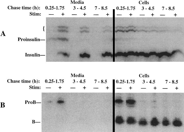
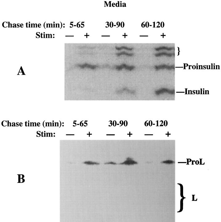
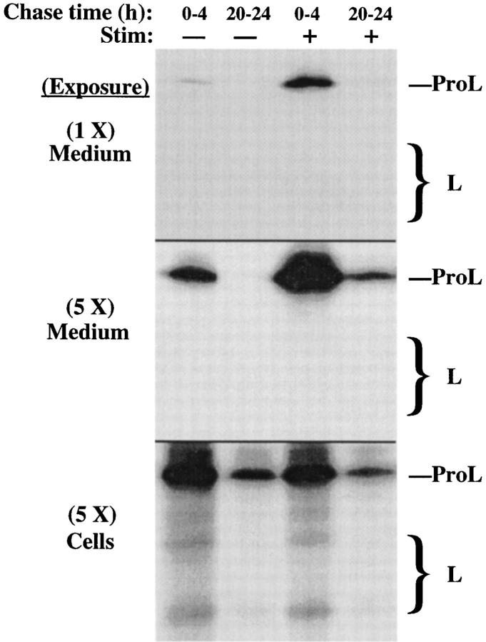
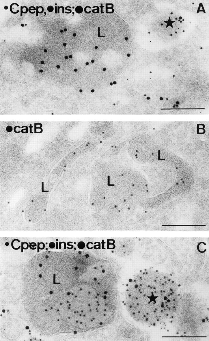
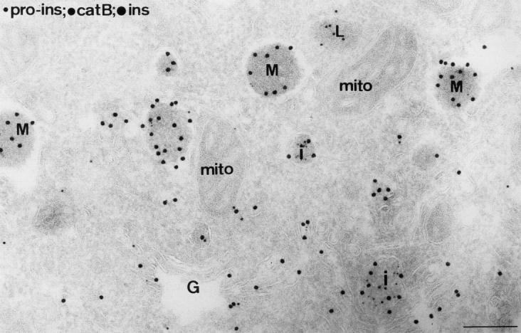
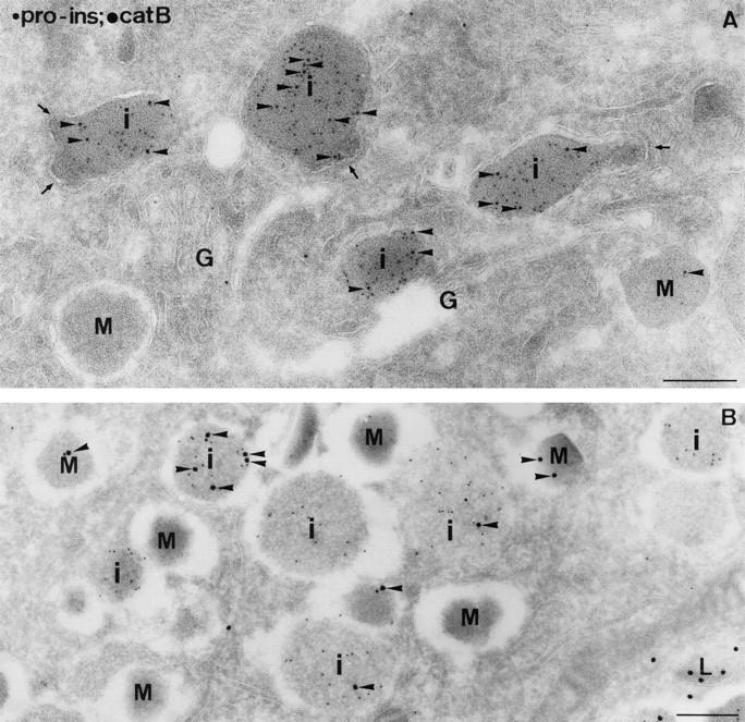
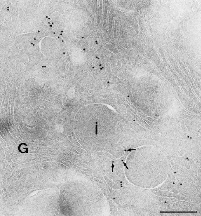
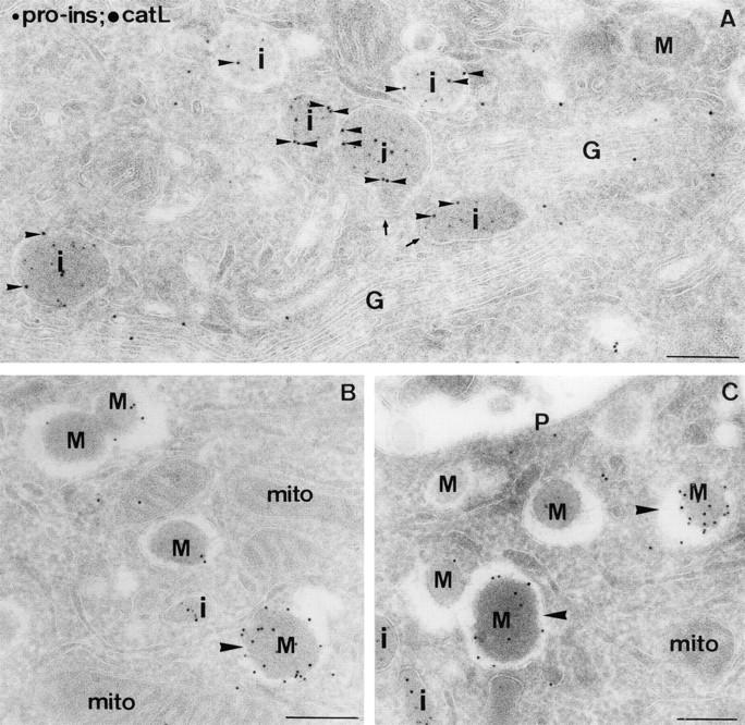
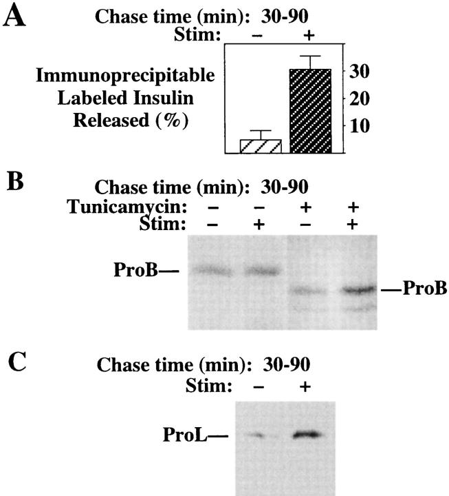
In cells specialized for secretory granule exocytosis, lysosomal hydrolases may enter the regulated secretory pathway. Using mouse pancreatic islets and the INS-1 beta-cell line as models, we have compared the itineraries of procathepsins L and B, two closely related members of the papain superfamily known to exhibit low and high affinity for mannose-6-phosphate receptors (MPRs), respectively. Interestingly, shortly after pulse labeling INS cells, a substantial fraction of both proenzymes exhibit regulated exocytosis. After several hours, much procathepsin L remains as precursor in a compartment that persists in its ability to undergo regulated exocytosis in parallel with insulin, while procathepsin B is efficiently converted to the mature form and can no longer be secreted. However, in islets from transgenic mice devoid of cation-dependent MPRs, the modest fraction of procathepsin B normally remaining within mature secretory granules is increased approximately fourfold. In normal mouse islets, immunoelectron microscopy established that both cathepsins are present in immature beta-granules, while immunolabeling for cathepsin L, but not B, persists in mature beta-granules. By contrast, in islets from normal male Sprague-Dawley rats, much of the proenzyme sorting appears to occur earlier, significantly diminishing the stimulus-dependent release of procathepsin B. Evidently, in the context of different systems, MPR-mediated sorting of lysosomal proenzymes occurs to a variable extent within the trans-Golgi network and is continued, as needed, within immature secretory granules. Lysosomal proenzymes that fail to be sorted at both sites remain as residents of mature secretory granules.


Figures

Figure 1
Figure 1
Stimulus-dependent release from INS cells of peptides immunoprecipitable with antiinsulin (A) or anti– cathepsin B (B). The cells, pulse labeled as described in Materials and Methods, were chased for up to 8.5 h. During different 90-min chase intervals, either stimulated (+) or unstimulated (−) secretion was collected (left), and the cells were then lysed. Equal fractions of media and cells were immunoprecipitated using anti-ProB, and one-tenth of these volumes were taken for precipitation with antiinsulin. The positions of proinsulin, insulin, presumptive proinsulin conversion intermediates (small bracket), ProB, and mature cathepsin B (B) are shown. Scanning densitometry of these gels led to the numerical data described in the text for this representative experiment (of three).
Figure 2
Figure 2
Stimulus-dependent release from INS cells of peptides immunoprecipitable with antiinsulin (A) or anti–cathepsin L (B). The cells were pulse labeled and chased as in Fig. 1, except that the stimulated (+) or unstimulated (−) medium collections began at 5 min of chase, were 60 min in duration, and were terminated at 2 h of chase. Only the medium is shown. While stimulated secretion of ProL is seen at all chase times, note the progression of processing from proinsulin to insulin in the regulated secretory pathway. Measurement of stimulus-dependent secretion during a 1-h period for L-containing peptides (∼16%) was in a similar range to that of insulin-containing peptides (∼21%) in this experiment. The positions of proinsulin, insulin, presumptive proinsulin conversion intermediates (small bracket), ProL, and bands comprising mature cathepsin L (M r ∼32,000 and ∼27,000, respectively, large bracket) are shown.
Figure 3
Figure 3
Inefficient arrival of newly synthesized ProL in lysosomes of INS cells. Cells pulse labeled as in Fig. 1 were chased to 3 h before a further 3-h collection of stimulated (+) or unstimulated (−) secretion was obtained and the cells analyzed by immunoprecipitation for lysosomal arrival of cathepsin L. Stimulus-dependent secretion contained ∼50% of all immunoprecipitable L-containing peptides found in the 3 h of collected medium plus cell lysate, while insulin secretion as measured by RIA averaged 53% during this period. The positions of ProL and bands comprising mature cathepsin L (large bracket) are shown.
Figure 4
Figure 4
A significant fraction of ProL in INS cells becomes entrapped in long-term storage in the regulated secretory pathway. The stimulated (+) and unstimulated (−) secretions from pulselabeled INS cells were collected either during the first 4 h after labeling or after a chase of 20 h to first allow all newly synthesized ProL molecules to reach their final targets. At the conclusion of each set, both media and cell lysates were analyzed by immunoprecipitation with anti–cathepsin L. Two exposures of the media, differing fivefold, are shown in the upper two panels. Note the disappearance of labeled forms of mature L at later chase times, and the persistent stimulus-dependent secretion of the precursor. The positions of ProL and bands comprising mature cathepsin L (large bracket) are shown.
Figure 5
Figure 5
Stimulus-dependent release from INS cells treated with tunicamycin. Untreated INS cells (left) or those pretreated with tunicamycin (right) were pulse labeled and chased for 3.5 h before 60-min collections of stimulated (+) or unstimulated (−) secretion, or the resulting cell lysates were analyzed by immunoprecipitation with antiinsulin (A) or anti– cathepsin L (B). For INS cells treated with tunicamycin, samples were intentionally double loaded to enhance the sensitivity of detection of mature cathepsin L forms. Note that after tunicamycin treatment, the amount of intracellular precursor increased disproportionately, as did the amount of stimulus-dependent secretion of ProL. The positions of proinsulin, insulin, presumptive proinsulin conversion intermediates (small bracket), glycosylated and unglycosylated ProL, and bands comprising glycosylated and unglycosylated mature cathepsin L (large brackets) are shown.
Figure 6
Figure 6
In β-cells of mouse pancreatic islets, lysosomes are morphologically distinct from secretory granules. (A) A typical lysosome (L) juxtaposed next to a mature secretory granule (star). The section was triple immunolabeled for C-peptide (5-nm gold), insulin (10-nm gold) and cathepsin B (15-nm gold). (B) Tubular lysosomes (L) immunopositive for cathepsin B (10-nm gold). (C) A crinophagic vacuole (L) juxtaposed next to a mature secretory granule (star). Triple immunolabeling was as in A. Consistent with previous reports (Orci et al., 1984b ; Halban et al., 1987), C-peptide labeling is absent from lysosomes, although insulin labeling may be present. Bars, 200 nm.
Figure 7
Figure 7
Ultrathin cryosection of the Golgi (G) region of a mouse islet β-cell, triple immunolabeled for proinsulin (5-nm gold), cathepsin B (10-nm gold), and insulin (15-nm gold). Insulin label was found over all compartments of the secretory pathway. Granule profiles with the presence of proinsulin label were used to define IGs (I), while those with insulin labeling only and a variable-sized halo between the content and surrounding membrane were considered mature granules (M). A lysosome (L) is seen to be labeled exclusively for cathepsin B, although additional cathepsin B label can be seen in IGs. mito, mitochondrion. Bar, 200 nm.
Figure 8
Figure 8
The occurrence of cathepsin B in IGs (i) of mouse islet β-cells. Ultrathin cryosections were double immunolabeled for proinsulin and cathepsin B. Cathepsin B label (arrowheads) was clearly present over proinsulin-positive IGs (i), whereas mature granules (M) were almost devoid of label. (A) Proinsulin (5-nm gold); cathepsin B (10-nm gold). Coated buds (arrows) were often observed on IGs, especially in the Golgi (G) region. Immunoreactive cathepsins are occasionally found directly in the buds, but, similar to reports from nonregulated secretory cells (Geuze et al., 1984, 1985; Ludwig et al., 1991), immunogold detection of hydrolase precursors tends to be impaired directly in the bud region. (B) Proinsulin (5-nm gold); cathepsin B (15-nm gold). L, lysosome. Bars, 200 nm.
Figure 9
Figure 9
Coated buds that form on IGs contain clathrin. Ultrathin cryosections were immunolabeled with an antibody to clathrin heavy chain. Labeling is present on numerous vesicular profiles at the trans-side of the Golgi complex (G) and on a membrane bud of an IG (i), marked with arrows. Bar, 200 nm.
Figure 10
Figure 10
The occurence of cathepsin L in IGs and mature granules of mouse islet β-cells. Ultrathin cryosections were double immunolabeled for proinsulin (5-nm gold) and cathepsin L (10-nm gold). (A) Overview of the Golgi (G) region. Cathepsin L labeling (small arrowheads) was clearly seen over proinsulin-positive IGs (i). The arrows point to coated buds on IGs. (B and C) Cathepsin L label was abundantly observed over both immature (i) and mature granule profiles (M), although some granules were more extensively labeled (large arrowheads) than others. mito, mitochondrion; P, plasmalemma. Bars, 200 nm.
Figure 11
Figure 11
Immunolabeling of cathepsins L and B in mature granules of mouse islet β-cells. (A and B) Ultrathin cryosections were immunolabeled for cathepsin L (10-nm gold) and insulin (15-nm gold), demonstrating the presence of cathepsin L labeling in mature β-granules. In B, two exocytotic profiles at the plasma membrane (P) are seen. (C) Triple labeling for insulin (5-nm gold), cathepsin L, (10-nm gold), and cathepsin B (15-nm gold) demonstrates a mature granule immunopositive for cathepsin L and immunonegative for cathepsin B (arrow). Occasional immunogold labeling of cathepsin B is indicated by arrowheads. Bars, 200 nm.
Figure 12
Figure 12
Stimulus-dependent exocytosis of mature β-granules releases an increased fraction of ProB from the islets of transgenic MPRdeficient mice. Islets from CD-MPR–deficient mice were pulse labeled and chased overnight in the absence of secretagogues to allow the labeled proinsulin and ProB to chase to their final destinations, before collecting sequential 30-min periods of unstimulated (−) and stimulated (+) secretion. See text for details. Secretion of labeled insulin and immunoprecipitable ProB were analyzed by SDS-PAGE and fluorography.
Figure 13
Figure 13
Unstimulated and stimulated secretion of newly synthesized insulin (A), ProB (B), and ProL (C) from pancreatic islets of normal male Sprague-Dawley rats. The islets, untreated or pretreated with tunicamycin, were pulse labeled for 30 min. During 30–90 min of chase, the islets were exposed either to unstimulated (−) or stimulated (+) conditions. Secretion of proinsulin + insulin was analyzed by immunoprecipitation as described (Arvan et al., 1991). Immunoprecipitable ProB and ProL were analyzed by SDS-PAGE and fluorography. After tunicamycin treatment, ProB was secreted as the unglycosylated form.

References

    1. Arvan P, Castle JD. Isolated secretion granules from parotid glands of chronically stimulated rats possess an alkaline internal pH and inward-directed H+pump activity. J Cell Biol. 1986;103:1257–1267. - PMC - PubMed
    1. Arvan P, Castle JD. Phasic release of newly synthesized secretory proteins in the unstimulated rat exocrine pancreas. J Cell Biol. 1987;104:243–252. - PMC - PubMed
    1. Arvan P, Castle JD. Protein sorting and secretion granule formation in regulated secretory cells. Trends Cell Biol. 1992;2:327–331. - PubMed
    1. Arvan P, Chang A. Constitutive protein secretion from the exocrine pancreas of fetal rats. J Biol Chem. 1987;262:3886–3890. - PubMed
    1. Arvan P, Lee J. Regulated and constitutive protein targeting can be distinguished by secretory polarity in thyroid epithelial cells. J Cell Biol. 1991;112:365–376. - PMC - PubMed

Publication types

MeSH terms