. 1997 May 19;137(4):835-45.
doi: 10.1083/jcb.137.4.835.

Characterization of the adaptor-related protein complex, AP-3

Affiliations

Characterization of the adaptor-related protein complex, AP-3

F Simpson et al. J Cell Biol. .

Abstract

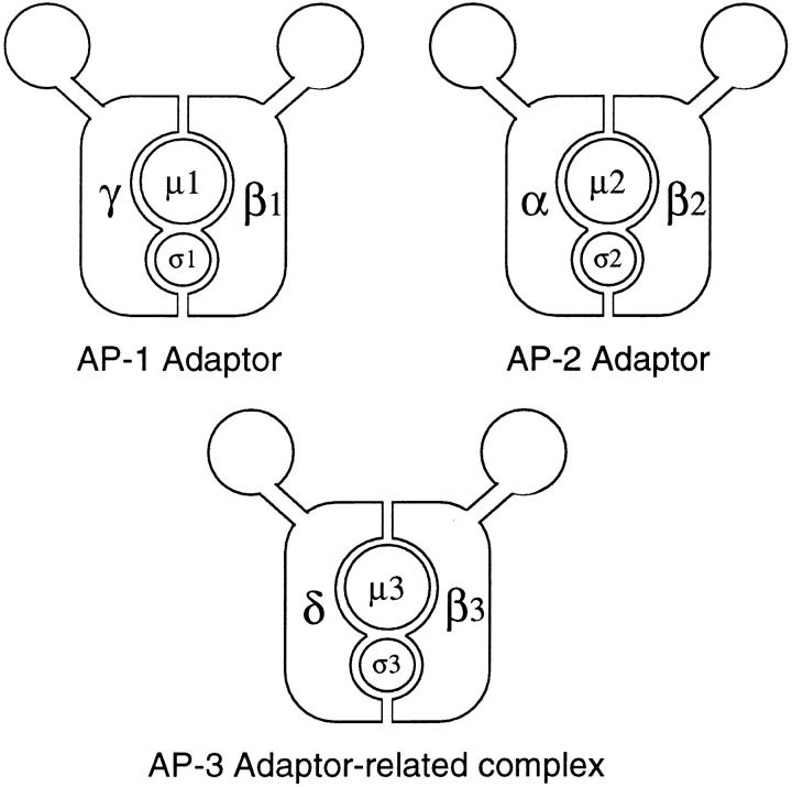
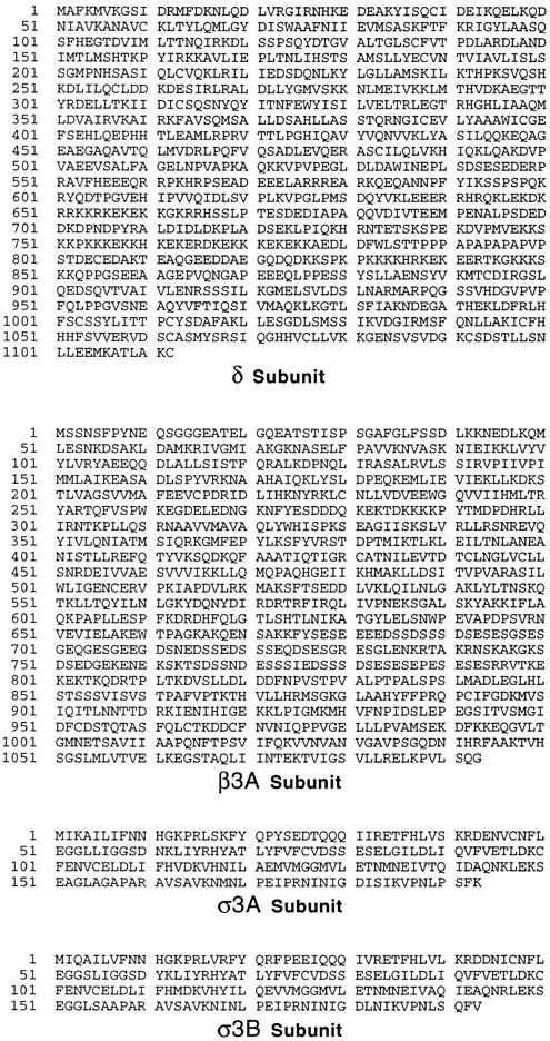
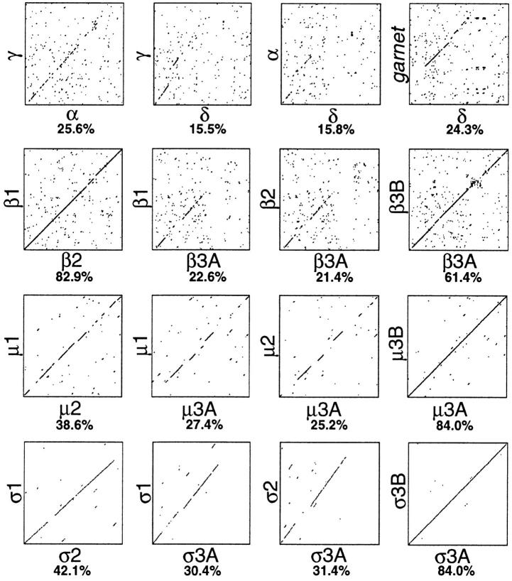
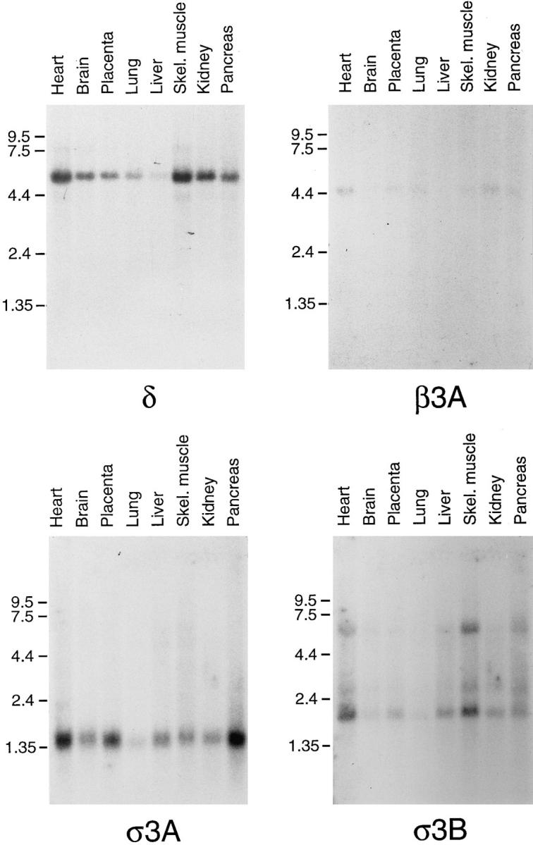
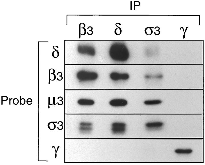
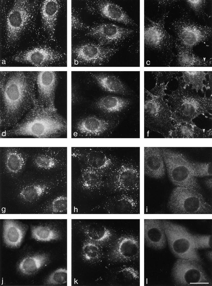
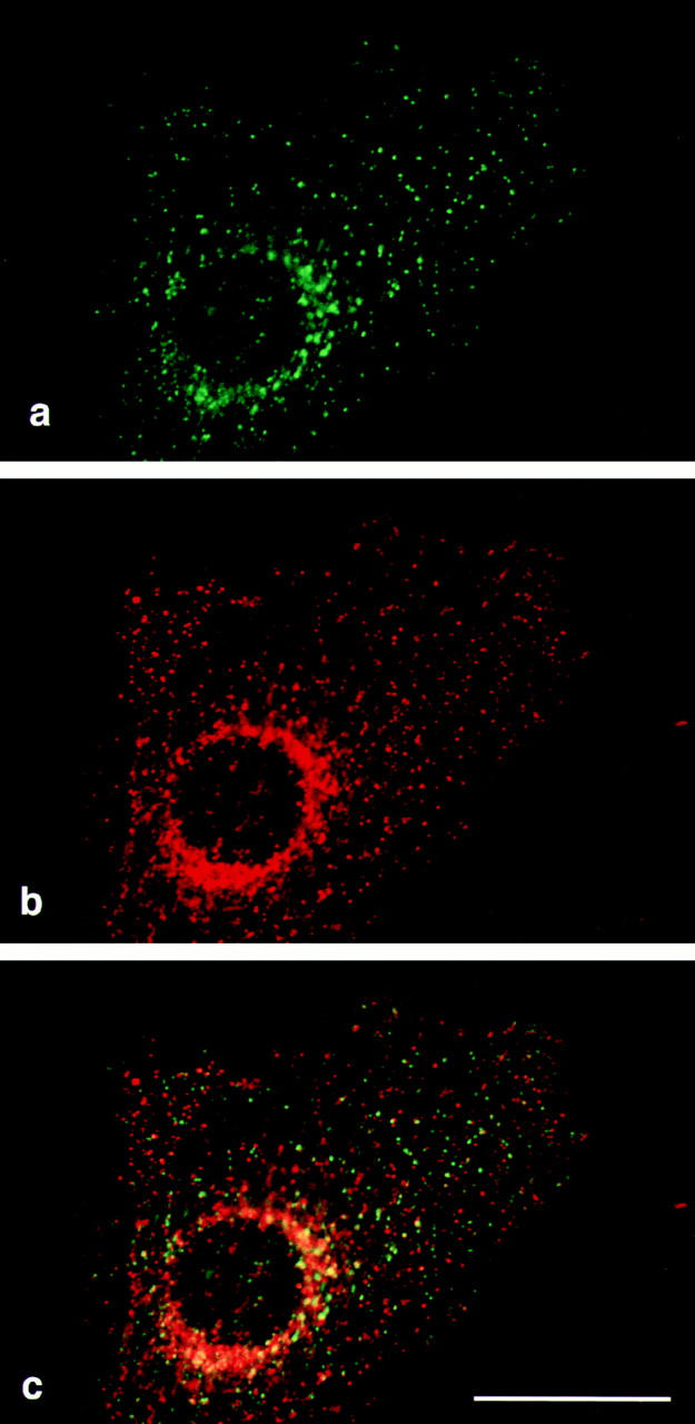
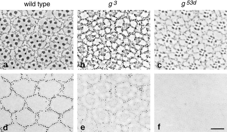
We have recently shown that two proteins related to two of the adaptor subunits of clathrincoated vesicles, p47 (mu3) and beta-NAP (beta3B), are part of an adaptor-like complex not associated with clathrin (Simpson, F., N.A. Bright, M.A. West, L.S. Newman, R.B. Darnell, and M.S. Robinson, 1996. J. Cell Biol. 133:749-760). In the present study we have searched the EST database and have identified, cloned, and sequenced a ubiquitously expressed homologue of beta-NAP, beta3A, as well as homologues of the alpha/gamma and sigma adaptor subunits, delta and sigma3, which are also ubiquitously expressed. Antibodies raised against recombinant delta and sigma3 show that they are the other two subunits of the adaptor-like complex. We are calling this complex AP-3, a name that has also been used for the neuronalspecific phosphoprotein AP180, but we feel that it is a more appropriate designation for an adaptor-related heterotetramer. Immunofluorescence using anti-delta antibodies reveals that the AP-3 complex is associated with the Golgi region of the cell as well as with more peripheral structures. These peripheral structures show only limited colocalization with endosomal markers and may correspond to a postTGN biosynthetic compartment. The delta subunit is closely related to the protein product of the Drosophila garnet gene, which when mutated results in reduced pigmentation of the eyes and other tissues. Because pigment granules are believed to be similar to lysosomes, this suggests either that the AP-3 complex may be directly involved in trafficking to lysosomes or alternatively that it may be involved in another pathway, but that missorting in that pathway may indirectly lead to defects in pigment granules.


Figures

Figure 1
Figure 1
Diagrams of the two conventional adaptor complexes, together with the adaptor-related complex, AP-3. The AP-1 complex is associated with the TGN, the AP-2 complex is associated with the plasma membrane, and the AP-3 complex also appears to be associated with the TGN as well as with more peripheral membranes. Each complex consists of four subunits, belonging to four different families. γ, α, and δ are related; β1 (β′), β2 (β), and β3 (β-NAP/β3B and β3A) are related; μ1 (AP47), μ2 (AP50), and μ3 (p47A/μ3A and p47B/μ3B) are related; and σ1 (AP19), σ2 (AP17), and σ3 (A and B) are related. EM studies of the AP-2 complex have revealed that it has a structure resembling a head flanked by two ears connected by flexible hinges (Heuser and Keen, 1988), and although such studies have not yet been carried out on AP-1 or AP-3, the sequence homologies suggest that all three complexes have a similar structure.
Figure 2
Figure 2
Sequences of novel components of the AP-3 complex. cDNAs encoding the four components were originally identified as ESTs encoding homologues of the known AP subunits. The complete sequences of the δ and β3A subunits were obtained by library screening. Both the cDNA and the protein sequence data are available from GenBank/EMBL/ DDBJ under accession numbers U91930 (δ), U91931 (β3A), U91932 (σ3A), and U91931 (σ3B).
Figure 3
Figure 3
Diagon plots comparing related proteins in the AP-1, AP-2, and AP-3 complexes. The sequences of all four types of subunits were compared using the “SIP” program (Staden, 1990), which was also used to calculate the percent identity. The δ subunit shows the least similarity with its counterparts in the AP-1 and AP-2 complexes, while the σ subunits (σ3A and σ3B) show the most. The protein product of the Drosophila garnet gene is also shown, compared with the δ subunit.
Figure 4
Figure 4
Expression patterns of δ, β3A, σ3A, and σ3B. Northern blots were probed with either oligonucleotides or cDNAs specific for each of the four sequences. All four genes are expressed ubiquitously. The relative weakness of the signal obtained with β3A probe is probably a consequence of the blot already having been probed several times.
Figure 5
Figure 5
Coimmunoprecipitation of AP-3 subunits. Pig brain cytosol was immunoprecipitated under nondenaturing conditions with affinity-purified polyclonal antibodies against β3B, δ, σ3 (crossreacting with both the A and B isoforms), and γ. Gels were blotted, and the appropriate region was cut out and probed with each of the above antibodies, as well as with anti-μ3 (which does not recognize the native complex). The four subunits of the AP-3 complex, δ, β3, μ3, and σ3, all coimmunoprecipitate; the γ subunit of the AP-1 complex does not coimmunoprecipitate with antibodies against the AP-3 components, and antibodies against γ do not bring down any of these components. The doublet labeled with anti-σ3 presumably corresponds to the A and B isoforms, one of which appears to be preferentially immunoprecipitated with this antibody.
Figure 6
Figure 6
Immunofluorescence localization of the δ subunit of the AP-3 complex. (a, b, d, and e) NRK cells were fixed with methanol/acetone and double labeled with anti-δ (a and b) and anti-transferrin receptor (d) or anti-lgp120 (e). Although all three antibodies show perinuclear labeling, many organelles in the cell are concentrated in this region, and there is little colocalization of the more peripheral elements. (c and f) Cells were allowed to endocytose rhodamine-conjugated wheat germ agglutinin (f) for 5 min before fixation in methanol/ acetone and were then labeled with anti-δ (c). Again, there is little colocalization of the more peripheral elements, although some structures do coincide (arrowheads). (g and j) NRK cells were fixed with paraformaldehyde and permeabilized with NP-40 and then double labeled with anti-δ (g) and antip200 (j), a putative TGN coat. Both antigens have a perinuclear distribution, but the fine details are different, indicating that they are not components of the same coat. (h, i, k, and l) MDBK cells were fixed with methanol/acetone, either with (i and l) or without (h and k) prior incubation for 2 min in 5 μg/ ml brefeldin A and then double labeled with anti-δ (h and i) and anti-γ (k and l). Both antigens are brefeldin A sensitive, and both have a similar perinuclear distribution in control cells, but little if any colocalization of more peripheral structures. Bar, 20 μm.
Figure 7
Figure 7
Confocal micrographs of an NRK cell double labeled for δ and clathrin. NRK cells were fixed with methanol/acetone and labeled with anti-δ (a, shown in green) and a monoclonal antibody against clathrin heavy chain (b, shown in red). The merged images (c) show numerous dots that are positive for δ but not for clathrin, and the limited amount of overlap is consistent with the two being found on the same membranes but different populations of coated buds. Bar, 20 μm.
Figure 8
Figure 8
Phenotype of Drosophila with mutations in the garnet gene, which encodes the δ subunit. Sections were cut from the eyes of both wild type (a and d) and mutant (b, c, e, and f) flies. Two alleles were examined: g3 (b and e) and g53d (c and f). a–c are phase contrast micrographs; d–f show brightfield views of the same images. Each ommatidium consists of photoreceptor cells surrounded by pigment cells. In wild-type flies the pigment granules in the pigment cells can be readily seen in brightfield as well as phase contrast micrographs (a and d). The granules are still visible in the g3 flies but contain less pigment and thus are less prominent when viewed by brightfield (b and e). They are essentially undetectable in the g53d flies (c and f). Bar, 10 μm.

References

    1. Ban J, Portetelle D, Altaner C, Horizon B, Milan D, Krchnak V, Burny A, Kettmann R. Isolation and characterization of a 2.3-kilobase-pair cDNA fragment encoding the binding domain of the bovine leukemia virus receptor. J Virol. 1993;67:1050–1057. - PMC - PubMed
    1. Bridges CB. Non-disjunction as proof of the chromosome theory of heredity. Genetics. 1916;1:1–52. - PMC - PubMed
    1. Brodsky FM. Clathrin structure characterized with monoclonal antibodies. I. Analysis of multiple antigenic sites. J Cell Biol. 1985;101:2047–2054. - PMC - PubMed
    1. Burkhardt JK, Wiebel FA, Hester S, Argon Y. The giant organelles in beige and Chediak-Higashi fibroblasts are derived from late endosomes and mature lysosomes. J Exp Med. 1993;178:1845–1856. - PMC - PubMed
    1. Chen MS, Obar RA, Schroeder CC, Austin TW, Poodry CA, Wadsworth SA, Vallee RB. Multiple forms of dynamin are encoded by shibire, a Drosophilagene involved in endocytosis. Nature (Lond) 1991;351:583–586. - PubMed

Publication types

MeSH terms

Associated data