. 1997 May 19;137(4):891-8.
doi: 10.1083/jcb.137.4.891.

Dictyostelium IQGAP-related protein specifically involved in the completion of cytokinesis

Affiliations

Dictyostelium IQGAP-related protein specifically involved in the completion of cytokinesis

H Adachi et al. J Cell Biol. .

Abstract

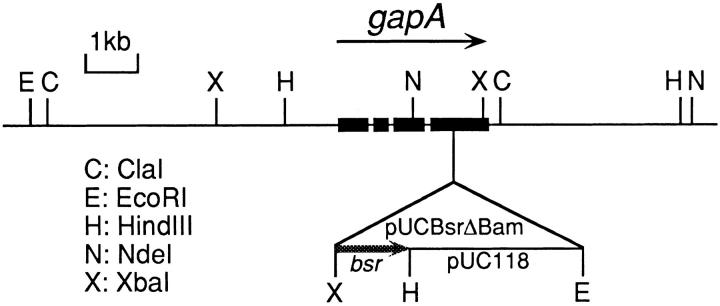
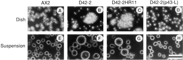
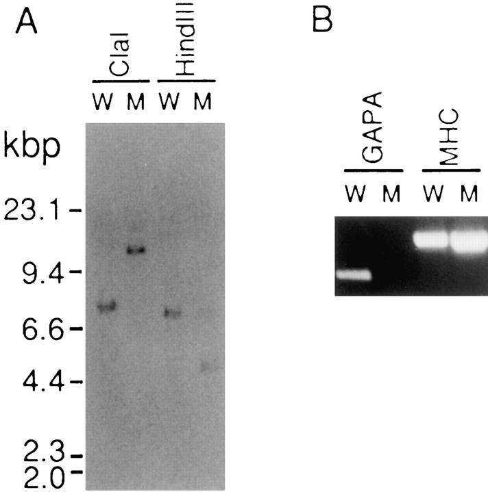
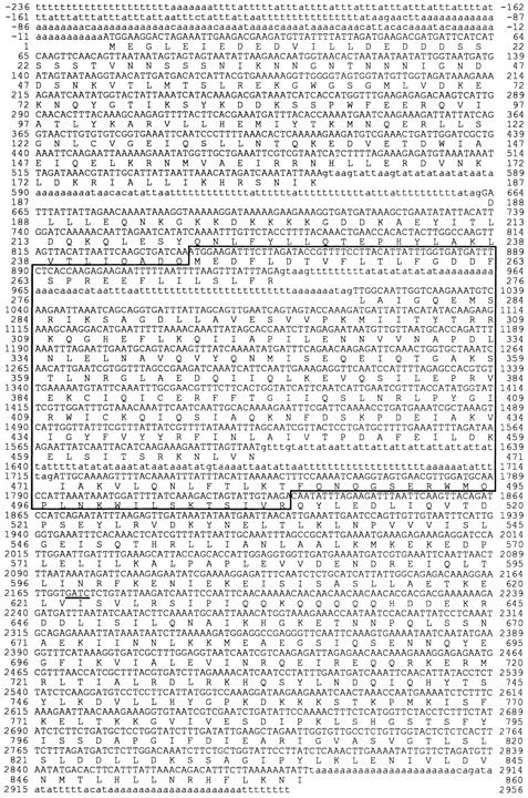
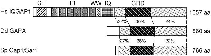
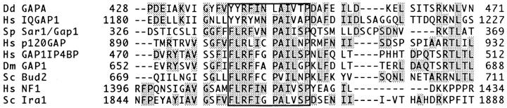
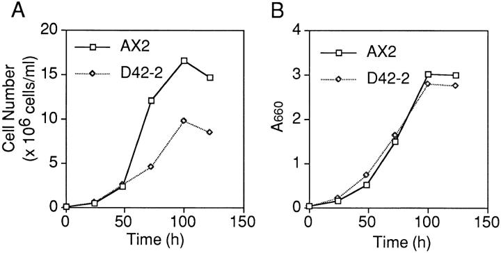
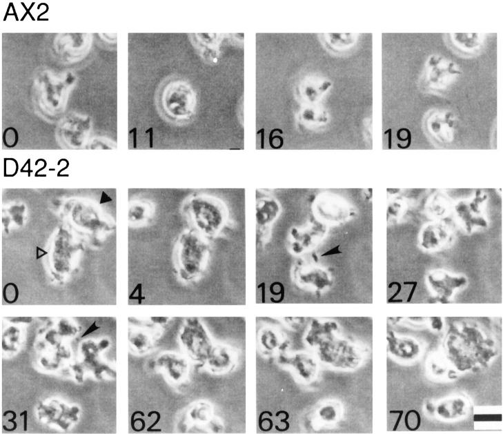
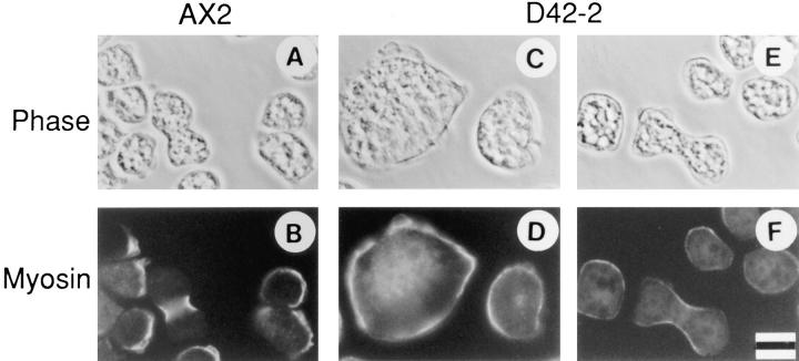
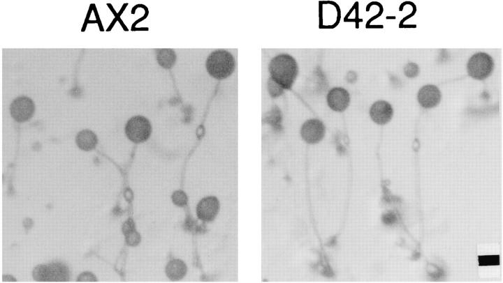
The gapA gene encoding a novel RasGTPase-activating protein (RasGAP)-related protein was found to be disrupted in a cytokinesis mutant of Dictyostelium that grows as giant and multinucleate cells in a dish culture. The predicted sequence of the GAPA protein showed considerable homology to those of Gap1/Sar1 from fission yeast and the COOH-terminal half of mammalian IQGAPs, the similarity extending beyond the RasGAP-related domain. In suspension culture, gapA- cells showed normal growth in terms of the increase in cell mass, but cytokinesis inefficiently occurred to produce spherical giant cells. Time-lapse recording of the dynamics of cell division in a dish culture revealed that, in the case of gapA- cells, cytokinesis was very frequently reversed at the step in which the midbody connecting the daughter cells should be severed. Earlier steps of cytokinesis in the gapA- cells seemed to be normal, since myosin II was accumulated at the cleavage furrow. Upon starvation, gapA- cells developed and formed fruiting bodies with viable spores, like the wild-type cells. These results indicate that the GAPA protein is specifically involved in the completion of cytokinesis. Recently, it was reported that IQGAPs are putative effectors for Rac and CDC42, members of the Rho family of GTPases, and participate in reorganization of the actin cytoskeleton. Thus, it is possible that Dictyostelium GAPA participates in the severing of the midbody by regulating the actin cytoskeleton through an interaction with a member of small GTPases.


Figures

Figure 1
Figure 1
Restriction map around the Dictyostelium gapA gene and the tag integration site of the REMI mutant, D42-2. Genomic DNA from the D42-2 strain was digested with the restriction enzymes indicated, and then analyzed by Southern blotting using linearized pUC118, a portion of the tag, as a probe. Information obtained on restriction analysis of the rescued plasmids is also included. The four exons of the gapA gene are indicated by thick lines. bsr, blasticidin S-resistance marker.
Figure 2
Figure 2
Size and shape of axenically growing Dictyostelium cells. (A–D) Cells were grown on coverslips, fixed, and then stained with 4′,6-diamidino-2-phenylindole. A phase-contrast image was laid over a fluorescent one. (E–H) Cells were grown in suspension (150 rpm), and living cells were observed by phase-contrast microscopy. (A and E) The parental strain, AX2; (B and F) the cytokinesis mutant, D42-2; (C and G) one of the mutants regenerated through homologous recombination, D42-2HR11; (D and H) the D42-2 strain harboring the p43-L plasmid carrying an intact gapA gene. Bar, 50 μm.
Figure 3
Figure 3
Southern blot (A) and reverse transcription–PCR (B) analyses of wild-type AX2 (W) and mutant D42-2 (M) cells. (A) 2.5 μg of genomic DNA digested with either ClaI or HindIII was loaded into each lane. The transferred DNA was probed with the full-length gapA cDNA. (B) DNA amplified with the same amount of total RNA and specific primers was loaded onto each lane. For details, see Materials and Methods.
Figure 4
Figure 4
DNA sequence of the Dictyostelium gapA gene and the deduced amino acid sequence of the GAPA protein. The coding DNA sequences (exons) are indicated by capital letters, and the 5′ and 3′ flanking sequences as well as introns are shown in lowercase letters. (Boxed region) RasGAP-related domain (GRD). In cytokinesis mutant D42-2, the tag was inserted into the underlined GATC sequence in the fourth exon. These sequence data are available from EMBL/GenBank/DDBJ under accession number D88027.
Figure 5
Figure 5
Schematic drawing of the similarity among the members of the IQGAP family. Identical residues (%) were calculated. CH, calponin homology domain; IR, IQGAP repeats; WW, WW domain; IQ, four repeats of the calmodulin-binding motif; GRD, RasGAP-related domain; Hs, Homo sapiens; Dd, Dictyostelium discoideum; Sp, Schizosaccharomyces pombe.
Figure 6
Figure 6
Amino acid sequence alignment of the most conserved region of RasGAP-related proteins. Identical residues are shaded. The invariant FLRXXXPAXXXP motifs are boxed. Dm, Drosophila melanogaster; Sc, Saccharomyces cerevisiae. GenBank accession numbers: Dd GAPA, D88027; Hs IQGAP1, D63875; Sp Sar1/Gap1, D10457; Hs p120GAP, M23379; Dm GAP1, M86655; Sc Bud2, L19162; Hs NF1, M89914; Sc Ira1, M24378. The sequence of GAP1IP4BP was taken from the original paper (Cullen et al., 1995).
Figure 7
Figure 7
Growth of Dictyostelium cells in axenic suspension. Wild-type AX2 and gapA D42-2 cells were diluted in fresh medium at 105 cells per ml, and then shaken at 22°C and 150 rpm. Their growth was monitored as the increase in cell number (A) or turbidity (B; absorbance at 660 nm) with the same culture.
Figure 8
Figure 8
Reversion of cytokinesis observed in gapA cells. Wildtype AX2 (upper frames) and mutant D42-2 (lower frames) cells were axenically grown on coverslips. Cell division was observed under a microscope connected to a time-lapse video recorder. The number in each frame indicates the time in min. In the lower frames (D42-2), the mutant cells (open and filled triangles) attempted to divide into two daughter cells. For the cell with the filled triangle, cytokinesis was reversed at the step where the two daughter cells were connected by a thin cytoplasmic bridge (midbody; arrowhead). In contrast, the cell with the open triangle completed cytokinesis like wild-type cells. Bar, 10 μm.
Figure 9
Figure 9
Localization of myosin II at the cleavage furrow. (A and B) AX2; (C–F) D42-2. Axenically growing cells were fixed by the agar-overlay method (Fukui et al., 1987). Myosin II was visualized by means of indirect immunofluorescence using DM2 mAbs against Dictyostelium myosin II (B, D, and F). (A, C, and E) Phase contrast. Bar, 10 μm.
Figure 10
Figure 10
Development of Dictyostelium cells upon starvation. (Left) AX2; (Right) D42-2. Vegetative cells were washed and then plated onto phosphate agar. The developed phenotype was observed after 24 h. Bar, 100 μm.

References

    1. Adachi H, Hasebe T, Yoshinaga K, Ohta T, Sutoh K. Isolation of Dictyostelium discoideumcytokinesis mutants by restriction enzyme-mediated integration of the blasticidin S resistance marker. Biochem Biophys Res Commun. 1994;205:1808–1814. - PubMed
    1. Amano M, Ito M, Kimura K, Fukata Y, Chihara K, Nakano T, Matsuura Y, Kaibuchi K. Phosphorylation and activation of myosin by rho-associated kinase (Rho-kinase) J Biol Chem. 1996a;271:20246–20249. - PubMed
    1. Amano M, Mukai H, Ono Y, Chihara K, Matsui T, Hamajima Y, Okawa K, Iwamatsu A, Kaibuchi K. Identification of a putative target for Rho as the serine-threonine kinase protein kinase N. Science (Wash DC) 1996b;271:648–650. - PubMed
    1. Bain G, Tsang A. Disruption of the gene encoding the p34/31 polypeptides affects growth and development of Dictyostelium discoideum. . Mol Gen Genet. 1991;226:59–64. - PubMed
    1. Brill S, Li S, Lyman CW, Church DM, Wasmuth JJ, Weissbach L, Bernards A, Snijders AJ. The ras GTPase-activating-protein-related human protein IQGAP2 harbors a potential actin binding domain and interacts with calmodulin and rho family GTPases. Mol Cell Biol. 1996;16:4869–4878. - PMC - PubMed

Publication types

Associated data