. 1997 May 19;137(4):899-908.
doi: 10.1083/jcb.137.4.899.

A glycine-rich RNA-binding protein mediating cold-inducible suppression of mammalian cell growth

Affiliations

A glycine-rich RNA-binding protein mediating cold-inducible suppression of mammalian cell growth

H Nishiyama et al. J Cell Biol. .

Abstract

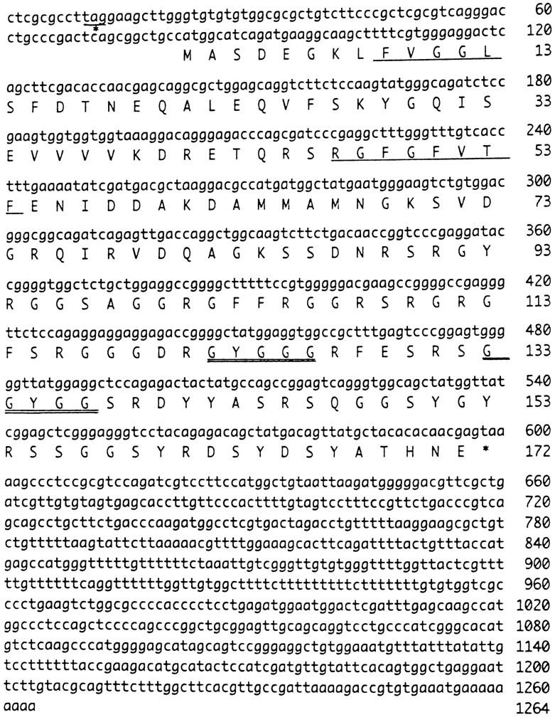
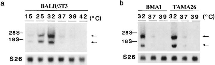
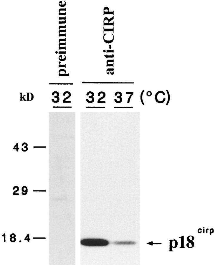
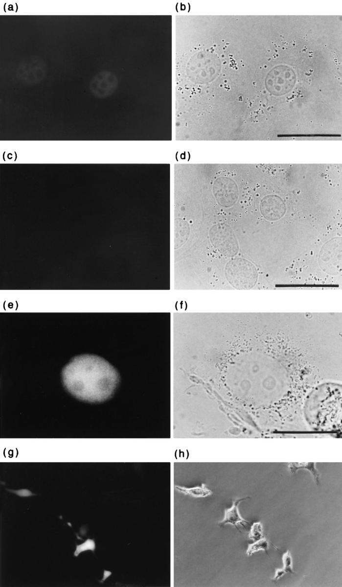
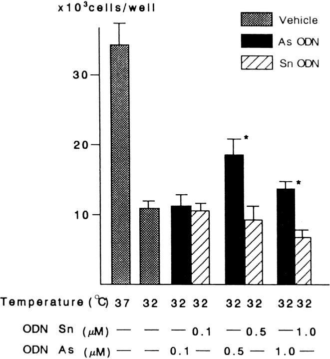
In response to low ambient temperature, mammalian cells as well as microorganisms change various physiological functions, but the molecular mechanisms underlying these adaptations are just beginning to be understood. We report here the isolation of a mouse cold-inducible RNA-binding protein (cirp) cDNA and investigation of its role in cold-stress response of mammalian cells. The cirp cDNA encoded an 18-kD protein consisting of an amino-terminal RNAbinding domain and a carboxyl-terminal glycine-rich domain and exhibited structural similarity to a class of stress-induced RNA-binding proteins found in plants. Immunofluorescence microscopy showed that CIRP was localized in the nucleoplasm of BALB/3T3 mouse fibroblasts. When the culture temperature was lowered from 37 to 32 degrees C, expression of CIRP was induced and growth of BALB/3T3 cells was impaired as compared with that at 37 degrees C. By suppressing the induction of CIRP with antisense oligodeoxynucleotides, this impairment was alleviated, while overexpression of CIRP resulted in impaired growth at 37 degrees C with prolongation of G1 phase of the cell cycle. These results indicate that CIRP plays an essential role in cold-induced growth suppression of mouse fibroblasts. Identification of CIRP may provide a clue to the regulatory mechanisms of cold responses in mammalian cells.


Figures

Figure 1
Figure 1
Cloning of cirp cDNA. Nucleotide and deduced amino acid sequences of the clone RNP78 containing cirp cDNA. Amino acid sequence is shown in single-letter code below the nucleotide sequence. The putative RNP motifs are underlined. GYGGG and GGYGG, well conserved among GRPs, are doubly underlined. Terminal codon is indicated by an asterisk. These sequence data are available from GenBank/EMBL/DDBJ under accession number D78135.
Figure 2
Figure 2
(a) Comparison of amino acid sequences in the RNA-binding domains of mouse CIRP, human RBM3 (Derry et al., 1995), B. napus BnGRP10 (Bergeron et al., 1993), A. thaliana Ccr1 and Ccr2 (Carpenter et al., 1994), and human hnRNP G (Soulard et al., 1993). The consensus sequence for the RNA-binding domain, as determined by Burd and Dreyfuss (1994), is also shown. Dots indicate amino acids identical to the CIRP sequence. The sequences, RNP1, RNP2, DRET, and MNGKXXDG, are boxed. (b) Structural comparison of CIRP with its related proteins. The length of each bar reflects the actual length of the sequences. The numbers in the black boxes indicate percent identity of amino acid sequence to CIRP in the CS-RBD (RNP motif). Note that the glycine-rich domain of cyanobacterium Anabaena variabilis RbpA1 (Sato, 1995) is smaller than that of CIRP.
Figure 3
Figure 3
(a) Southern blot analysis of EcoRI-digested DNA from nine eukaryotic species (zoo blot analysis). DNAs were from human (lane 1), monkey (lane 2), rat (lane 3), mouse (lane 4), dog (lane 5), cow (lane 6), rabbit (lane 7), chicken (lane 8), and yeast (lane 9). The coding region of cirp cDNA was 32P-labeled and used as a probe. Autoradiography was done overnight (lanes 1–7) or for 3 d (lanes 8–9) at −80°C. Mobilities of molecular size markers, HindIII-digested λ phage DNA, are indicated on the left. (b) Southern blot analysis of mouse genomic DNA. 20 μg of liver DNAs extracted from either ddy/std mouse (lanes 1, 3, and 5) or C57BL/6 mouse (lanes 2, 4, and 6) were digested with one of the three restriction enzymes, EcoRI (lanes 1 and 2), PvuII (lanes 3 and 4), and KpnI (lanes 5 and 6), electrophoresed in 0.8% agarose gel, and transferred to a nylon membrane. The coding region of cirp cDNA was 32P-labeled and used as a probe. Autoradiography was done overnight.
Figure 4
Figure 4
RNA-binding property of CIRP. (a) Bacterially expressed GST protein and GST–CIRP fusion protein demonstrated by Coomassie blue staining. (b) Northwestern blots showing binding of GST and GST–CIRP proteins to radiolabeled ribonucleotide homopolymers, poly(A), poly(C), poly(G), and poly(U), at different salt concentrations as indicated.
Figure 5
Figure 5
Temperature-dependent expression of cirp mRNA. Northern blot analysis of total RNAs from mouse cell lines, BALB/3T3 (a) and BMA1 and TAMA26 (b), harvested 24 h after the indicated temperature shift. The positions of 18S and 28S ribosomal RNAs are indicated on the left. As a control for the amount of RNA loaded, the filter was rehybridized with a mouse S26 ribosomal protein cDNA probe (lower).
Figure 6
Figure 6
Western blot analysis of BALB/3T3 cell lysates using an anti-CIRP polyclonal antibody. BALB/3T3 cells were harvested 24 h after the indicated temperature shift. 10 μg of samples was separated by 14% SDS-PAGE. Note specific recognition of an 18-kD protein by the anti-CIRP antibody. Mobilities of coelectrophoresed molecular size markers are indicated on the left.
Figure 7
Figure 7
Kinetics of p18cirp induction in response to cold stress. BALB/3T3 cells were harvested at indicated times (hour) after a temperature shift from 37 to 32°C. Mobilities of coelectrophoresed molecular size markers are indicated on the left.
Figure 8
Figure 8
Localization of CIRP. Immunofluorescence microscopy of BALB/3T3 cells cultured at 32°C and stained with an anti-CIRP polyclonal antibody (a) or preimmune serum (c). The bound antibody was detected by an FITC-conjugated second antibody. Fluorescence microscopy of COS-7 cells expressing GFP–CIRP fusion protein (e) or GFP (g). Light (b, d, and f) or phasecontrast (h) microscopic images of the field of view identical to a, c, e, and g, respectively. Bars, 20 μm.
Figure 9
Figure 9
Effects of antisense (As) ODN on the cold-induction of p18cirp. BALB/3T3 cells were incubated at the indicated temperatures for 12 h in the presence of vehicle alone, As, or sense (Sn) ODNs (0.5 μM). Note partial suppression of the cold-induced p18cirp expression in the presence of antisense ODN.
Figure 10
Figure 10
Effects of antisense (As) ODN on the cold-induced suppression of cell growth. Cell numbers were determined after 2 d of culture at the indicated temperature in the presence or absence of indicated concentrations of ODN. The results are expressed as the mean ± SEM. *Statistically different from controls by unpaired Student's t test (P < 0.02).
Figure 11
Figure 11
Expression of p18cirp in representative transfectants. BALB/3T3 cells, cloned BALB/3T3 cells transfected with cirpexpression vector DNA (BAC-Sn3), or vector DNA (BAC-C1) were cultured for 12 h at the indicated temperature and analyzed by Western blotting.

References

    1. Aloia RC, Raison JK. Membrane function in mammalian hibernation. Biochim Biophys Acta. 1989;988:123–146. - PubMed
    1. Bandziulis RJ, Swanson MS, Dreyfuss G. RNA-binding proteins as developmental regulators. Genes Dev. 1989;3:431–437. - PubMed
    1. Bergeron D, Beauseigle D, Bellemare G. Sequence and expression of a gene encoding a protein with RNA-binding and glycine-rich domains in Brassica napus. . Biochim Biophys Acta. 1993;1216:123–125. - PubMed
    1. Burd CG, Dreyfuss G. Conserved structures and diversity of functions of RNA-binding proteins. Science (Wash DC) 1994;265:615–621. - PubMed
    1. Burdon RH. Temperature and animal cell protein synthesis. Symp Soc Exp Biol. 1987;41:113–133. - PubMed

Publication types

MeSH terms

Associated data